Tiny packages help nerve cells boost local protein-making by about 30%
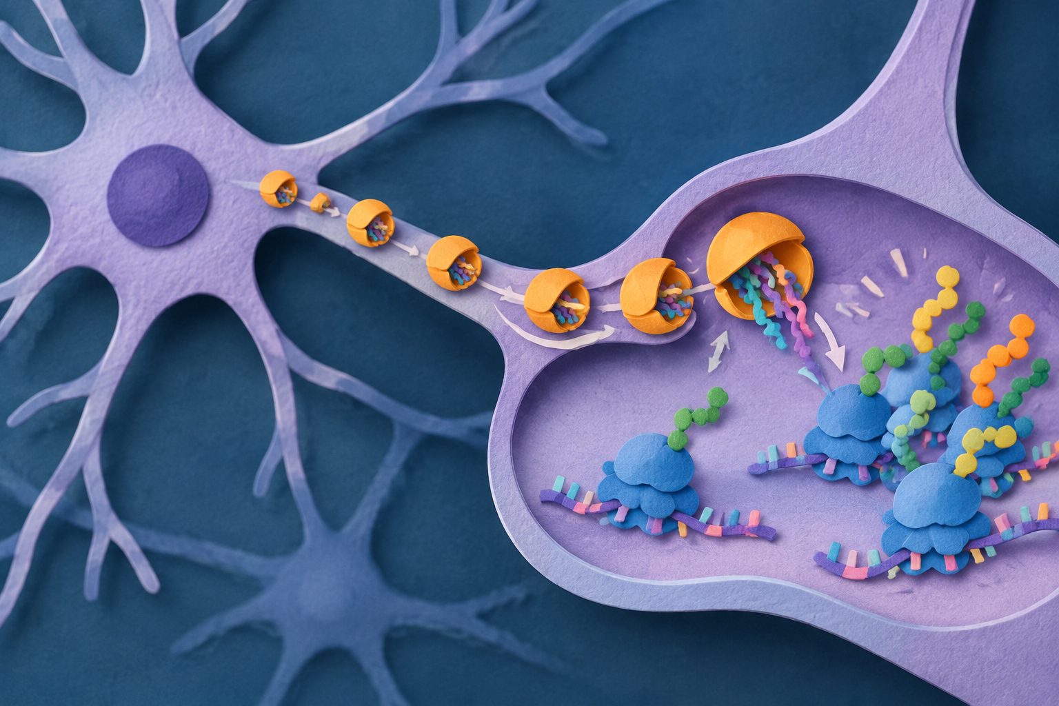
Ribosome-carrying vesicles move through axons and dendrites, helping neurons respond to stimulation by making proteins where they are needed.
Editorial illustration — not from the study.
Researchers used several forms of microscopy, including live imaging and electron microscopy, to study ribosome-associated vesicles in neuronal processes. They found these vesicles in rodent neurons and in an imaged human cortex sample, with ribosomes arranged on their surfaces in patterns consistent with active protein production.
Chemically induced long-term potentiation changed vesicle behavior differently in dendrites and axons. Dendrites accumulated more stationary vesicles, while axonal vesicles moved faster. The stimulation also increased the share of vesicles carrying ribosomes, expanding the pool available for local protein production.
How the vesicles work
The researchers identified endoplasmic-reticulum-derived vesicles marked by the KDEL protein signal, some of which were decorated with 80S ribosomes. Electron microscopy showed that the ribosomes formed ordered arrangements compatible with polysomes, groups of ribosomes that can make protein from the same messenger RNA.
In live imaging, about 66% of the vesicles were moving and 34% were stationary. Their speeds ranged from 0.2 to 3.2 micrometres per second, with a mean of 0.86 micrometres per second. After chemically induced long-term potentiation, dendrites gained more stationary vesicles, while axonal vesicles accelerated without a clear increase in their number.
At single-messenger-RNA resolution, translation sites associated with a ribosome-carrying vesicle had about 30% greater newly made-protein signal amplitude than periods without such an association. Together, the results indicate that these vesicles help deliver ribosomes to sites of activity-dependent local translation.
Why local protein-making matters
Protein production in axons and dendrites allows neurons to respond locally rather than relying only on proteins made in the cell body and transported over longer distances. The findings provide a mechanism for how ribosomes can reach these distant parts of a neuron and how neuronal activity can adjust that delivery differently in axons and dendrites.
By linking vesicle movement with local protein production, the study offers a framework for understanding how neurons control protein synthesis in both space and time during synaptic plasticity.
Evidence and caveats
The study combines cryo-electron tomography, volume electron microscopy and live-cell imaging. Experiments were performed mainly in cultured rodent neurons, with additional imaging of human cortex and mouse sensory neurons. The imaging provides structural and temporal evidence linking ribosome-carrying vesicles with local translation, while the roughly 30% comparison concerns translation-signal amplitude at associated versus unassociated sites.
The stimulation experiments used a chemical model of long-term potentiation, so they do not by themselves show how the vesicles behave during every form of neuronal activity or in an intact behaving animal. The reported findings also do not establish effects on behavior, disease or long-term synaptic function.
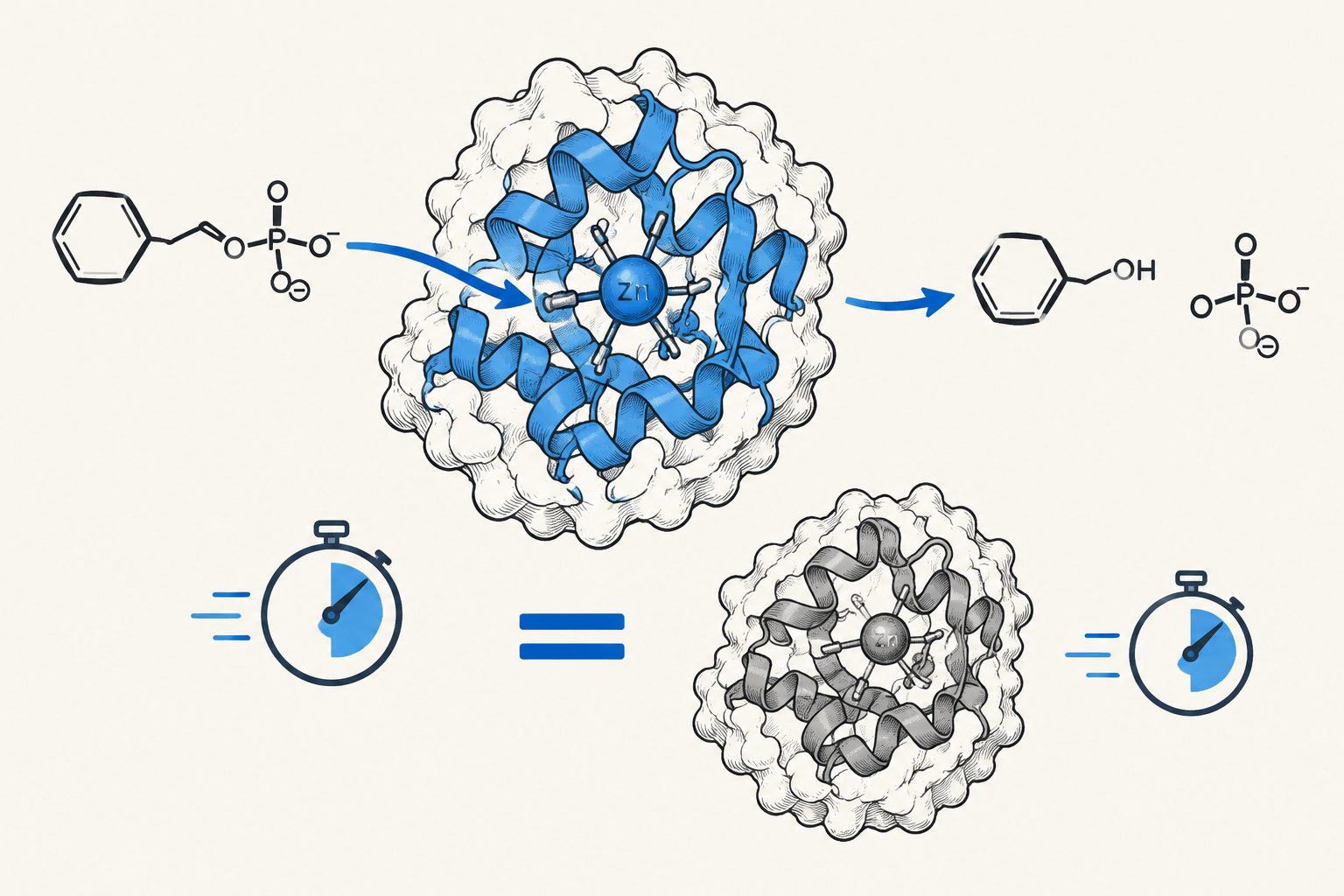
Researchers designed a zinc-containing enzyme from scratch, without a natural template, catalytic motif or evolutionary data. The enzyme showed activity against both phosphomonoesters and phosphodiesters, with rate enhancement comparable to characterized natural phosphatases.
A study of illicit alcohol in two Ugandan cities found that alcohol strength was inconsistent and sometimes under-declared, reaching 44.2%. Some spirits contained methanol above safe limits, while opaque beers showed bacterial contamination and many production and retail sites had poor sanitation.
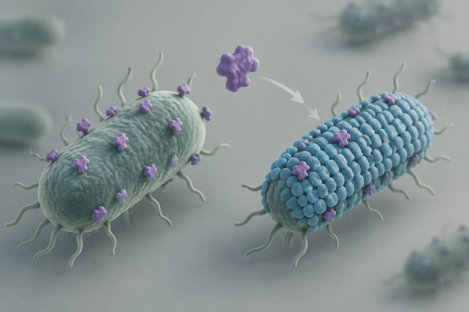
Researchers found that bacterial surface-layer proteins can evolve through modular changes and gene transfer, producing different outer structures. In closely related bacteria with the same type of surface layer, only pathogens showed resistance to the antimicrobial molecules LL-37 and lysozyme.