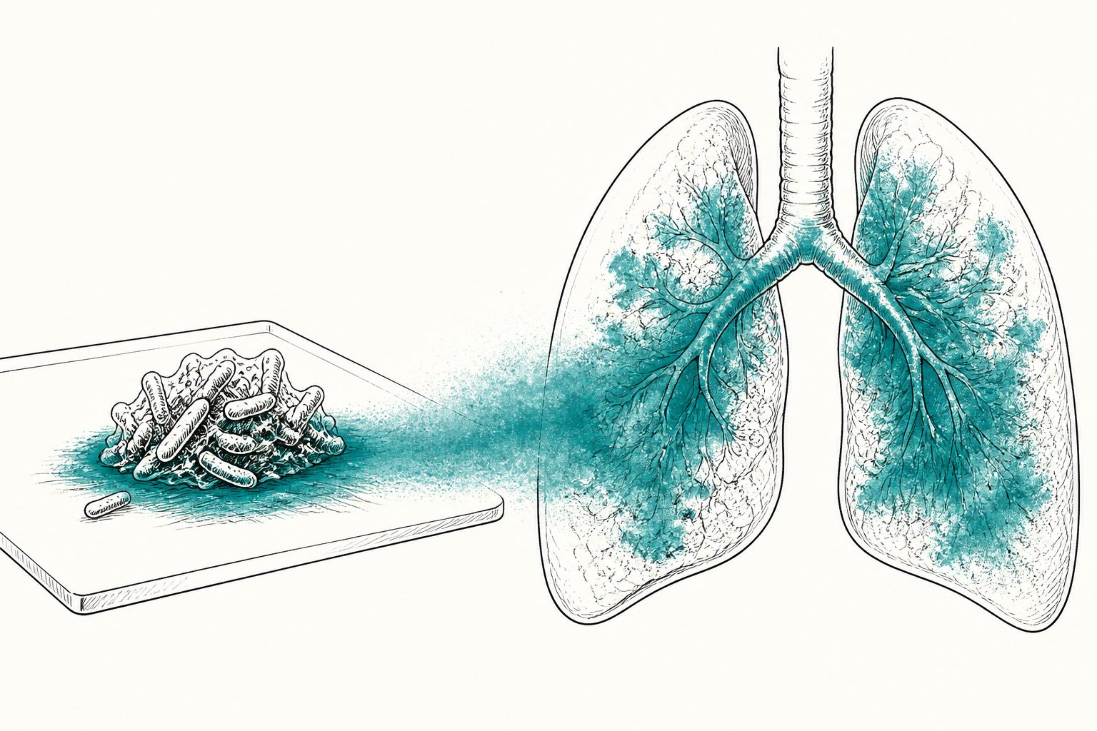
Chemical mapping found deeper pyocyanin presence in mouse lung explants than in biofilms grown on a synthetic surface.
Researchers grew Pseudomonas aeruginosa biofilms on polydimethylsiloxane, a synthetic material used as a laboratory model for biomedical devices, and on pieces of healthy mouse lung. They used matrix-assisted laser desorption/ionization mass spectrometry imaging to map molecules across the biofilms and surrounding tissue without adding labels.
Both models showed a broadly conserved set of molecules involved in bacterial communication, biofilm structure and metabolism. The lung-explant biofilms differed in the distribution of phenazines, especially pyocyanin, which was more prominent and penetrated deeply into the lung tissue.
What the imaging showed
The imaging method mapped several molecular families within the biofilms, including quorum-sensing molecules, phenazines and rhamnolipids. The overall molecular profile was conserved between the synthetic-surface and lung-explant models, but their spatial patterns differed. In particular, biofilms formed on lung explants showed increased pyocyanin production, with pyocyanin spreading into the lung tissue. The study also optimized sample preparation, embedding materials and sectioning approaches to preserve biofilm structure and tissue integrity during imaging.
// Source
Biofilm · 2026 · DOI: 10.1016/j.bioflm.2026.100401
Authors: Marie Droniou, C. Le Guillou, Hung Le, Christophe Arnoult, Teddy Grandjean, Philippe Gosset, Pascal Cosette, Isabelle Schmitz
Institutions: Inserm, Centre National de la Recherche Scientifique, Université de Lille, Centre Hospitalier Universitaire de Lille, Normandie Université, Université de Rouen Normandie, Institut National des Sciences Appliquées Rouen Normandie, Polymères, Biopolymères, Surfaces, Institut Pasteur de Lille, Center for Infection and Immunity of Lille, Communication Bactérienne et Stratégie Anti-infectieuses