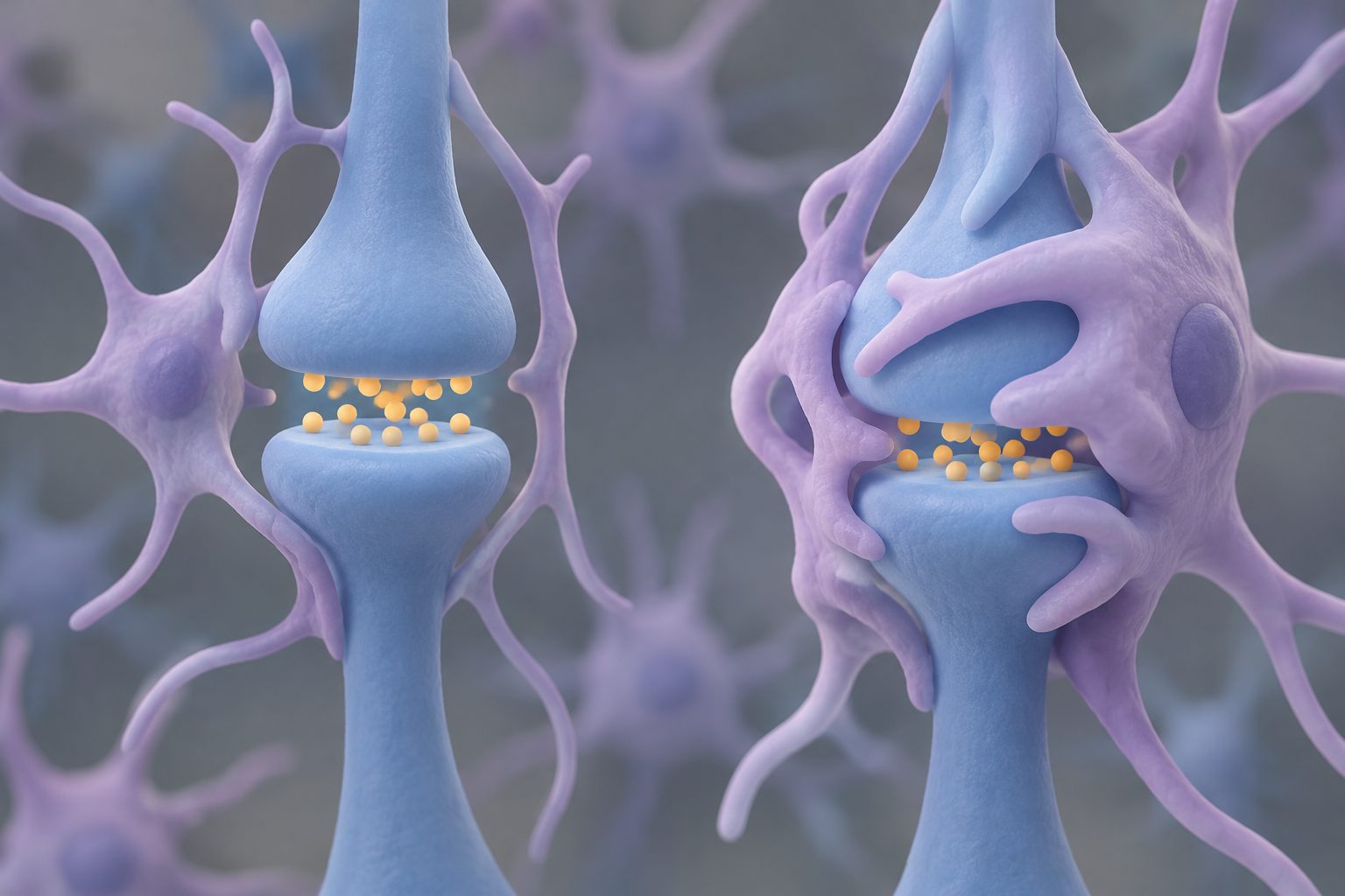
A study in young mice links the loss of FMRP to excessive synapse removal by microglia and astrocytes.
Researchers examined male mice lacking FMRP, the protein missing in Fragile X syndrome, at postnatal day 7. In the retinogeniculate pathway—a developing visual circuit used to study synapse removal—the mice had smaller synapses and faster separation of inputs from the two eyes.
Gene activity measurements showed coordinated changes in microglia, astrocytes and neurons in pathways linked to synapse removal. Computer modeling suggested stronger signaling from astrocytes to microglia, while a cell-based assay found that both types of glial cells engulfed more synaptic material in the lateral geniculate nucleus of the Fragile X mice.
Evidence and caveats
The study combined measurements of synapse structure, eye-specific wiring, cell-specific gene activity, computational estimates of cell-to-cell signaling, preliminary lipid profiling and a glial engulfment assay. The experiments were performed in male Fmr1 knockout mice at postnatal day 7, so the findings come from an early developmental mouse model rather than from people with Fragile X syndrome. The lipid results were preliminary, and the researchers say the functional importance of those changes remains uncertain. The computational signaling results are predictions, while the engulfment assay provides supporting cellular evidence; the study does not establish that these changes cause human Fragile X symptoms or show that altering them would improve outcomes.
// Source
Scientific Reports · 2026 · DOI: 10.1038/s41598-026-75819-z
Authors: Lindsey Starr, Melissa Lee, Amy Vo, Maia Weisenhaus, Lucas Cheadle, Archana Yadav, Fahad Paryani, Mimi Shirasu‐Hiza, Vilas Menon, Carol Ann Mason
Institutions: Columbia University, Harvard University, Columbia University Irving Medical Center, Cold Spring Harbor Laboratory, Mortimer B. Zuckerman Mind Brain Behavior Institute