A map of the mouse spinal cord shows cell changes linked to nerve pain
Researchers charted where spinal cord cell types occur and found altered gene activity and signaling patterns in mice with neuropathic pain.
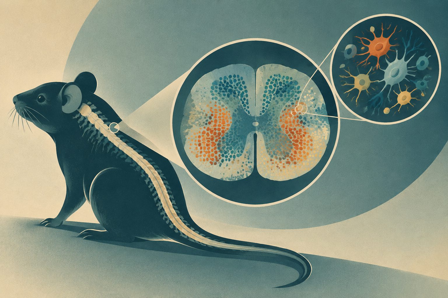
Editorial illustration — not from the study.
Researchers used MERFISH, an imaging method that measures gene activity in individual cells while preserving their locations, to examine cervical, thoracic, lumbar and sacral sections of adult mouse spinal cords. The study analyzed 146,621 high-quality cells from five male and five female mice and identified 60 transcriptionally distinct cell subtypes, along with non-neuronal cell types.
The resulting map showed that many neuronal subtypes varied in abundance across spinal levels and occupied distinct layers. Cells in the dorsal horn also displayed spatial patterns consistent with possible signaling between neighboring cell types. In a mouse model of neuropathic pain, several neuronal subtypes had changed gene-expression states and predicted cell-to-cell communication patterns.
What the map revealed
The researchers identified 14 broad cell classes and 60 transcriptionally distinct subtypes in the adult mouse spinal cord. These included excitatory and inhibitory neurons, motor neurons, cholinergic neurons and several non-neuronal cell types.
Neuronal subtypes were distributed differently across cervical, thoracic, lumbar and sacral levels, and some showed sex-specific differences in abundance. Many subtypes were positioned close to particular other subtypes, suggesting possible region-specific connections. In the dorsal horn, the researchers identified a range of predicted signaling interactions based on the locations of cells and the presence of matching ligand-receptor or neuropeptide-related molecules.
In mice with neuropathic pain, several neuronal subtypes showed altered transcriptomic states—changes in their measured gene activity—as well as altered predicted cell-to-cell communication. The excerpts do not specify which subtypes or signaling pairs changed, or how these changes relate to the severity or duration of pain.
Why the map matters
The spinal cord handles sensory information and helps coordinate motor and autonomic functions, but the organization of its many cell types across body-related spinal levels is not fully understood. This map links cell identity with location and provides a resource for studying how regional and sex-specific spinal circuits operate.
The pain-related results point to specific neuronal populations and communication patterns that may be involved in neuropathic pain. Because the study identifies molecular and spatial associations rather than testing treatments, its main value is as a guide for future work on the circuits involved in healthy function and disease.
Evidence and caveats
The study used MERFISH to measure a selected panel of 500 genes in individual cells while recording their positions in tissue. The data came from cervical, thoracic, lumbar and sacral spinal cord sections from five male and five female mice, with 146,621 high-quality cells collected across six independent technical replicates.
Spatial proximity does not establish that two cell types are functionally connected, and the predicted ligand-receptor interactions do not demonstrate that signaling occurs. The researchers state that connectomic and electrophysiological studies are needed to test functional connectivity. The findings also come from mice and from a model of neuropathic pain, so their relevance to human spinal-cord biology or human pain is not established by this study. The 500-gene panel provides targeted spatial information rather than a complete measurement of every gene.
// Source
Proceedings of the National Academy of Sciences · 2026 · DOI: 10.1073/pnas.2623177123
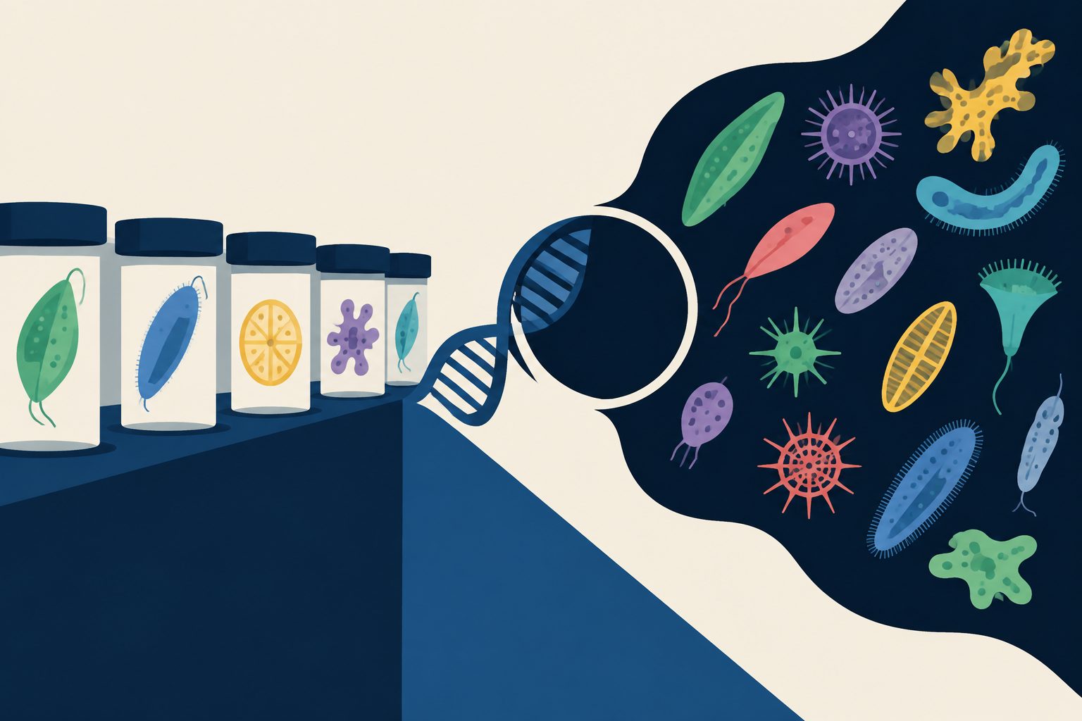
A survey of more than 30,000 metagenomes and other DNA datasets found extensive protist diversity with no described representative in reference collections. The analysis identified 13 deeply divergent candidate lineages and predicted links between some of them and bacteria or other eukaryotes.
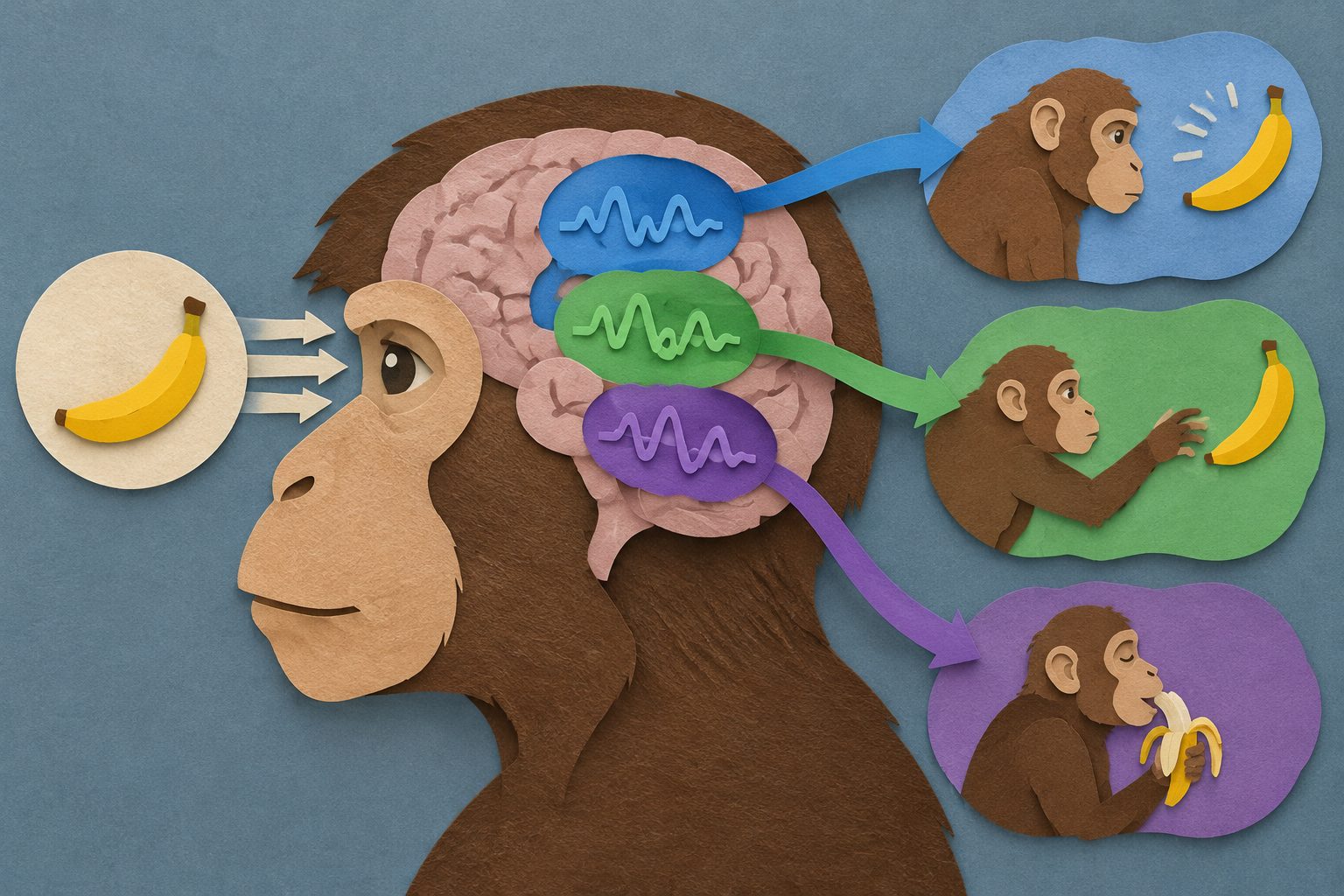
In a task combining visual motion and reward, monkeys’ brain regions represented the same information in different formats. A model predicted that stimulating those regions would have different behavioral effects, which experiments then observed.
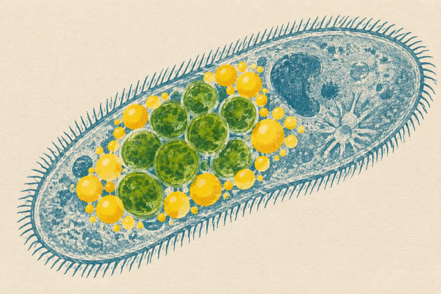
A study of the ciliate Paramecium bursaria found that its lipid droplets are reorganized when green algae live inside it. The droplets gathered near the algae, and chemical disruption of lipid metabolism reduced the algae’s numbers, pointing to a role in maintaining the symbiosis.