Researchers compare CT methods for tracking lung disease and lung-function decline
In 92 people with fibrosing interstitial lung disease, visual and automated CT measures showed similar associations with changes in breathing tests.
Low evidenceHuman studySome caution advised
Medical disclaimer: This article summarizes research findings and is for informational purposes only. It is not medical advice.
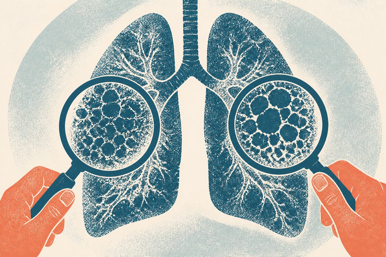
Editorial illustration — not from the study.
Researchers reviewed paired thin-section CT scans and pulmonary function tests from 92 people with fibrosing interstitial lung disease at one medical center. The tests were performed a median of 44.5 months apart. They compared a visual score based on five CT features with automated three-dimensional CT volume-histogram measurements.
The visual score was highly consistent between readers. Changes in both the visual score and automated measurements were associated with changes in forced vital capacity and carbon-monoxide diffusion, which are measures of lung function. The automated measures had similar associations to the visual score and showed good ability to identify radiological or physiological progression under the study’s definition, but these findings are associations from a retrospective study.
What was studied
The researchers examined whether an automated three-dimensional CT volume-histogram analysis could assess progression of fibrosing interstitial lung disease as consistently as visual scoring based on the 2022 radiological progressive pulmonary fibrosis criteria. They retrospectively studied 92 people at a single center who had paired thin-section CT scans and pulmonary function tests, with a median interval of 44.5 months. Five guideline-defined CT features were visually scored on selected axial images, and changes in skewness, kurtosis, entropy, and total lung volume were calculated from the CT scans. The researchers then compared these changes with changes in percent-predicted forced vital capacity and diffusing capacity for carbon monoxide, and assessed how well the measures identified progression as defined by the guideline.
What researchers observed
The change in the visual CT score had excellent agreement between observers, with intraclass correlation coefficients of 0.94–0.96. Changes in the visual score were strongly associated with changes in the automated CT measures: correlations were −0.78 for skewness, −0.74 for kurtosis, and 0.73 for entropy.
Automated CT measures showed substantial associations with changes in forced vital capacity, with correlation values ranging from 0.61 to 0.64 in absolute terms. The visual score had a correlation of −0.60 with forced vital capacity change. Associations with diffusing capacity for carbon monoxide were moderate: absolute correlation values ranged from 0.48 to 0.58 for the reported measures. Change in total lung volume was also associated with changes in forced vital capacity and diffusing capacity. All reported correlations were statistically significant, with p<0.001. The measures had area-under-the-curve values of 0.76–0.89 for identifying radiological and/or physiological progression under the study definition. These results indicate association and measurement performance, not causation or proof that automated analysis improves patient care.
Who this is relevant to
These findings may be most relevant to people with fibrosing interstitial lung disease who are evaluated with serial CT scans and pulmonary function tests in settings similar to the single center studied. They do not establish that the results apply to all forms of interstitial lung disease, people with different disease severity, or centers using different scanners and analysis methods. They also do not show that automated CT analysis is suitable for individual clinical decisions or that it improves outcomes.
Why this matters
Assessing change in fibrosing interstitial lung disease can involve detailed visual review of CT scans and lung-function testing. This study suggests that automated CT measurements may provide information similar to a reproducible visual score when researchers assess imaging changes and lung-function decline. However, the study did not test whether using automated analysis changes clinical decisions, treatment, quality of life, survival, or other patient outcomes.
Limitations & evidence assessment
The study was retrospective, conducted at one center, and included only 92 people, so selection and other biases are possible. Its observational design means the reported relationships cannot establish cause and effect. The abstract does not provide enough information about the participants’ specific diagnoses, how they were selected, whether the CT analyses were validated at other centers, or how missing data and potential confounding factors were handled. The median follow-up interval was long, but the abstract does not describe the timing and consistency of all measurements in detail. The findings were based on selected CT sections for visual scoring and on the study’s definition of progression, so they may not generalize to every patient or clinical setting.
Why this evidence level: This was a single-center retrospective observational study of 92 people, so it can show associations but cannot establish that one CT method causes better outcomes or is superior in clinical care. The sample was relatively small, and the abstract provides limited information about participant selection and other possible sources of bias.
Evidence levels are editorial estimates derived from study metadata — they are not clinical appraisals.
// Source
Japanese Journal of Radiology · 2026 · DOI: 10.1007/s11604-026-02070-x
A prospective, multicenter phase II trial examined whether removing only the treated tumor area through the anus could preserve the rectum in people with locally advanced rectal cancer who responded well to chemoradiotherapy. Among 80 participants, three-year disease-free survival was 84.8%, and local recurrence occurred in 5.0% during the reported follow-up. Because everyone received local excision and there was no comparison group, the findings do not establish that this approach is as effective as more extensive surgery.
A systematic review and meta-analysis examined how silver diamine fluoride affects human dental pulp cells in laboratory studies. Across seven in vitro studies, higher concentrations were associated with lower cell viability, but these findings do not show how the material affects dental pulp in people.
This systematic review examined human studies of long-term quality of life and depressive symptoms after fracture-related infection, a serious infection involving a broken bone. Across 15 studies, physical quality-of-life scores appeared lower than United States population norms, while evidence about mental quality of life and depressive symptoms was less consistent. The researchers said differences among studies and generally limited study quality prevent definitive conclusions.