New analysis finds silver diamine fluoride can harm dental pulp cells in lab studies
The review found that cell damage increased as the concentration rose, but the results came from laboratory models rather than patients.
Very low evidenceReviewInterpret with caution
Medical disclaimer: This article summarizes research findings and is for informational purposes only. It is not medical advice.
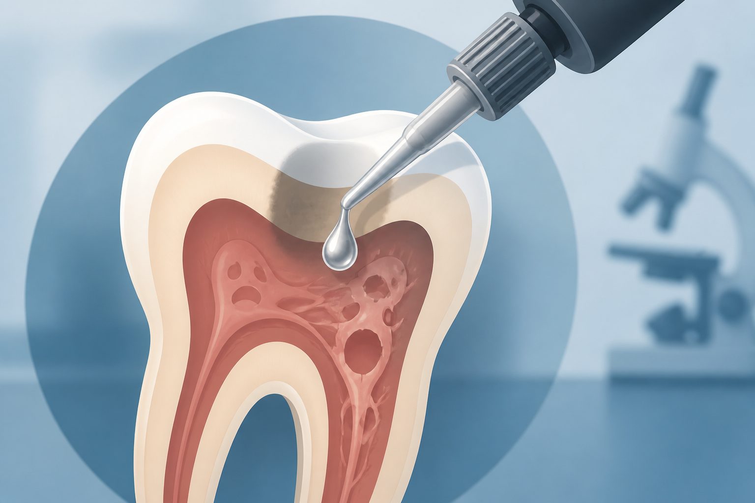
Editorial illustration — not from the study.
Researchers combined results from seven laboratory studies published from 2022 to 2024. The studies exposed human dental pulp cells to different concentrations of silver diamine fluoride and measured cell viability and other biological effects.
The review reported little cytotoxicity at very low concentrations, declines in cell viability at 0.001% to 0.005%, and severe toxicity at concentrations of 0.008% or higher. The authors said conventional concentrations used clinically may pose a risk near the dental pulp, but the review did not directly study patients or clinical outcomes.
What the review covered
The researchers conducted a PRISMA-compliant systematic review and meta-analysis of in vitro and ex vivo research on silver diamine fluoride and human dental pulp cells. Searches covered PubMed, Scopus, Cochrane CENTRAL, and Google Scholar without date restrictions. Seven in vitro studies from 2022 to 2024 were included, and cell-viability results were combined using random-effects statistical models. The available information does not state the total number of cells, laboratory experiments, or specific cell donors.
Key conclusions
The qualitative findings described a dose-dependent pattern: higher concentrations of silver diamine fluoride were generally associated with greater cytotoxicity, meaning more damage or loss of viability in the tested cells. The review reported minimal cytotoxicity at 0.00001% to 0.0005%, with a pooled effect of 0.99 and a 95% confidence interval of 0.97 to 1.00. At 0.001% to 0.005%, the pooled effect was 0.73, with a 95% confidence interval of 0.46 to 0.93. At concentrations of 0.008% or higher, the pooled effect was 0.14, with a 95% confidence interval of 0.00 to 0.41. Most included studies reported half-maximal inhibitory concentration values at or below 0.001%. These are laboratory cell results and do not by themselves establish injury, safety, or a particular clinical outcome in people.
Where this may apply
These findings apply most directly to human dental pulp cells studied under laboratory conditions. They do not directly apply to patients, to all dental procedures, or to the clinical effects of silver diamine fluoride in people. The review can inform further research about possible cellular toxicity, but it cannot by itself determine clinical safety or outcomes.
What this could mean
The findings are relevant to questions about how silver diamine fluoride might interact with dental pulp cells when exposure occurs in laboratory conditions. They suggest that concentration is an important factor in the tested cell models. However, laboratory exposure does not reproduce all conditions in a tooth or a patient, so the results cannot establish whether near-pulpal use causes pulpal injury in humans or identify a clinically safe concentration.
Limitations & evidence assessment
The evidence was based on only seven in vitro studies, so the overall amount of research was small. Laboratory cell models do not capture the full structure of a tooth, protective tissues, exposure duration, or other factors present in patients, and the studies did not report clinical outcomes. The abstract provides limited detail about differences among the studies, including cell sources, exposure conditions, and the statistical variability between studies. It also does not provide enough information to assess possible publication bias or determine how well the laboratory concentrations correspond to exposure in actual dental procedures.
Why this evidence level: This is a systematic review and meta-analysis, but it combined only seven laboratory studies using human dental pulp cells. Laboratory cell findings can help identify possible biological effects but do not establish what happens in patients or determine clinical safety.
Evidence levels are editorial estimates derived from study metadata — they are not clinical appraisals.
// Source
Journal of Oral Biology and Craniofacial Research · 2026 · DOI: 10.1016/j.jobcr.2026.101548
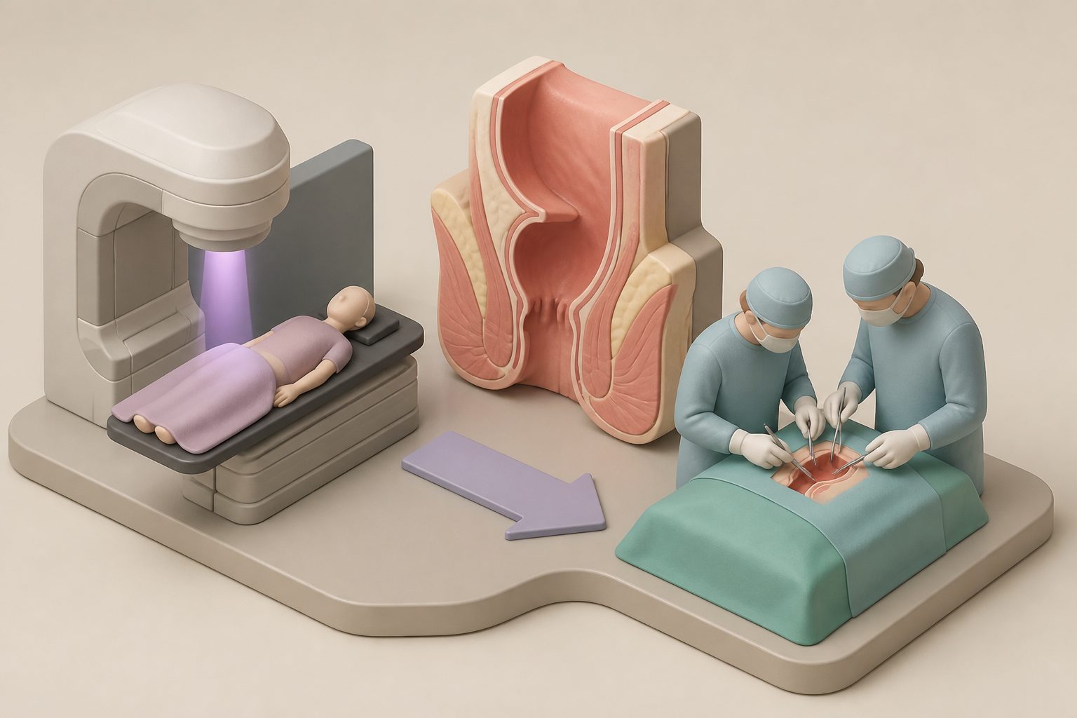
A prospective, multicenter phase II trial examined whether removing only the treated tumor area through the anus could preserve the rectum in people with locally advanced rectal cancer who responded well to chemoradiotherapy. Among 80 participants, three-year disease-free survival was 84.8%, and local recurrence occurred in 5.0% during the reported follow-up. Because everyone received local excision and there was no comparison group, the findings do not establish that this approach is as effective as more extensive surgery.
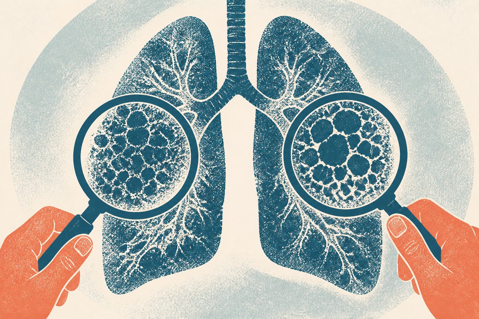
This retrospective human study compared a visual CT scoring method with an automated computer analysis for tracking fibrosing interstitial lung disease. Among 92 people followed with CT scans and lung-function tests, both approaches were associated with changes in lung function and signs of disease progression. The study does not show that either method improves treatment or patient outcomes.
This systematic review examined human studies of long-term quality of life and depressive symptoms after fracture-related infection, a serious infection involving a broken bone. Across 15 studies, physical quality-of-life scores appeared lower than United States population norms, while evidence about mental quality of life and depressive symptoms was less consistent. The researchers said differences among studies and generally limited study quality prevent definitive conclusions.