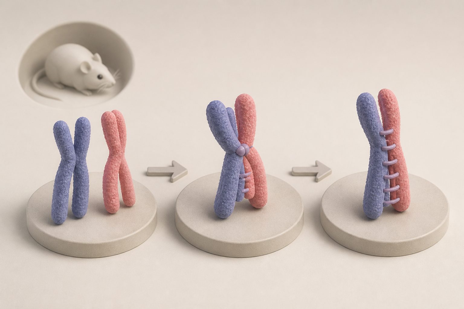
Live imaging shows how DNA recombination and chromosome-joining structures guide the process in developing mouse sperm cells.
Using a fluorescent labeling system and 3D time-lapse imaging, the researchers watched specific chromosome sites in living mouse seminiferous tubule tissue. They found that homologous chromosomes first approached from an average separation of 4.8 micrometres, reached an intermediate distance of 1.7 micrometres, and then came within 0.7 micrometres of one another as a chromosome-joining structure formed between them.
Studies of mice carrying changes in recombination-related genes, together with DNA imaging and chromosome staining, linked the first rapid transition to DNA double-strand breaks and early recombination events. The final close pairing depended on later crossover intermediates and the SYCP1-containing synaptonemal complex.
How the chromosomes paired
Homologous chromosomes did not pair in one continuous step. They moved through three measurable stages: widely separated, at an average distance of 4.8 micrometres; intermediate, at 1.7 micrometres; and closely paired, at 0.7 micrometres. The transition from the first stage to the second was relatively rapid.
The researchers observed rapid chromosome movements led by telomeres, the chromosome ends. Their analyses linked the shift from wide separation to intermediate pairing with DNA double-strand breaks and early recombination events. Close pairing correlated with formation of the synaptonemal complex, a structure that joins paired chromosomes, and required crossover maturation intermediates and SYCP1.
Evidence and caveats
The evidence comes from real-time 3D imaging of fluorescently labeled chromosome regions in live mouse seminiferous tubule tissue, supported by experiments in recombination mutants, DNA fluorescence imaging, and staining of chromosome spreads. The results show associations between particular recombination steps and pairing transitions, while the mutant analyses support the stated requirements for those transitions.
The fluorescent labeling system has limitations. Integrating large DNA-repeat arrays could alter local chromosome structure or nearby gene regulation, and the fluorescent binding protein could affect chromosome movement at the labeled site. Uneven labeling, limited access to the tagged DNA, delivery efficiency, and optical clarity can also reduce signal quality and the timing or spatial resolution of the imaging.
// Source
Nature Communications · 2026 · DOI: 10.1038/s41467-026-78036-4
Authors: Luciana P. de Almeida, V. Meg Butler, Leandra Marín, Katarzyna P Nowak, Marcin Ostoja-Helczynski, Cole Ragsdale, Monika K. Kawecka, Ky'ara Carr, Paula Elaine Cohen, Michael E. Dresser, Sean M. Burgess, Elizabeth H. Finn, Roberto J. Pezza
Institutions: Cornell University, University of California, Davis, University of Oklahoma Health Sciences Center, Oklahoma Medical Research Foundation, Oklahoma City University