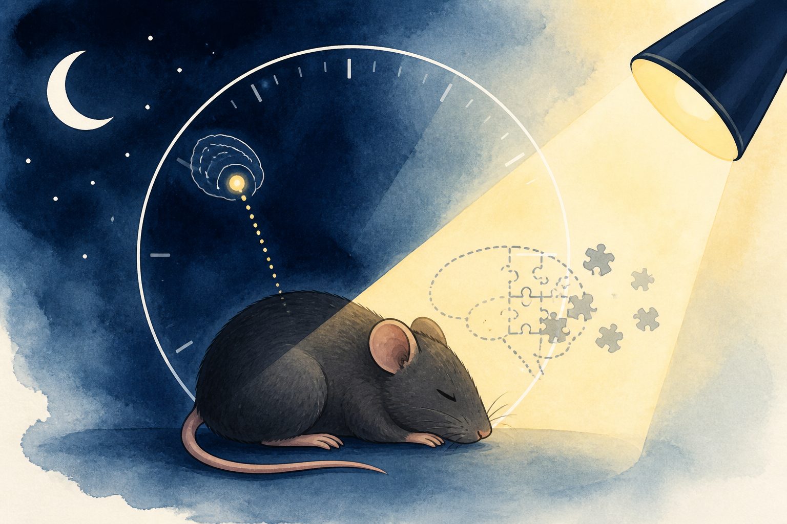
Six days of mistimed light altered brain clock activity and impaired long-term object recognition in male mice.
Researchers exposed male C57BL/6J mice to light during their circadian rest phase for six days, together with enforced wakefulness. The mice later showed impaired long-term object recognition memory, but their short-delay spatial memory was not impaired.
The mistimed light changed the organization and signaling of the brain’s central clock and reduced rhythmic activity of clock genes in the hippocampus, a region involved in memory. It also altered time-dependent synaptic gene activity and reduced a form of synaptic strengthening in the CA1 region. A local hippocampal treatment with dexamethasone restored day–night differences in clock-gene activity and preserved memory under the light exposure.
What the light changed
Rest-phase light exposure, given for six days, impaired long-term object recognition memory in male C57BL/6J mice but did not impair short-delay spatial memory. The exposure altered the organization and output signaling of the suprachiasmatic nucleus, the brain’s central circadian clock, and reduced the normal rhythmic activity of clock genes in the hippocampus.
In the hippocampus, mistimed light disrupted phase-dependent synaptic gene expression in dentate gyrus and CA1 neurons and reduced long-term potentiation in CA1, a cellular process linked to learning and memory. Mice kept awake without light exposure retained object recognition memory and CA1 clock-gene expression, indicating that mistimed light was required for the reported effects in this experiment. Local hippocampal dexamethasone treatment restored day–night differences in hippocampal clock-gene expression and preserved memory during rest-phase light exposure.
Evidence and caveats
The study used behavioral testing, single-nucleus RNA sequencing, and ex vivo electrophysiology in male C57BL/6J mice. A comparison group that experienced enforced wakefulness without light exposure helped test whether mistimed light was necessary for the observed changes, and local dexamethasone treatment provided further evidence that hippocampal clock regulation was involved.
The evidence comes from mice, not humans, and the experiment used male animals of one strain. The exposure combined mistimed light with enforced wakefulness, so the findings apply to this controlled model rather than to every form of late-night light exposure. The abstract does not report the study’s sample sizes.
// Source
Communications Biology · 2026 · DOI: 10.1038/s42003-026-10920-7
Authors: Xuebing Zhang, Changwei Yang, Wenyi Jin, Nan Zhang, Liang Zhang, Xingqi MENG, Yool Lee, Jin Young Kim
Institutions: Fujian Medical University, City University of Hong Kong, Washington State University, Washington State University Spokane