The markers helped cryo-electron microscopy reveal how drugs reshape signaling receptors without attached signaling partners.
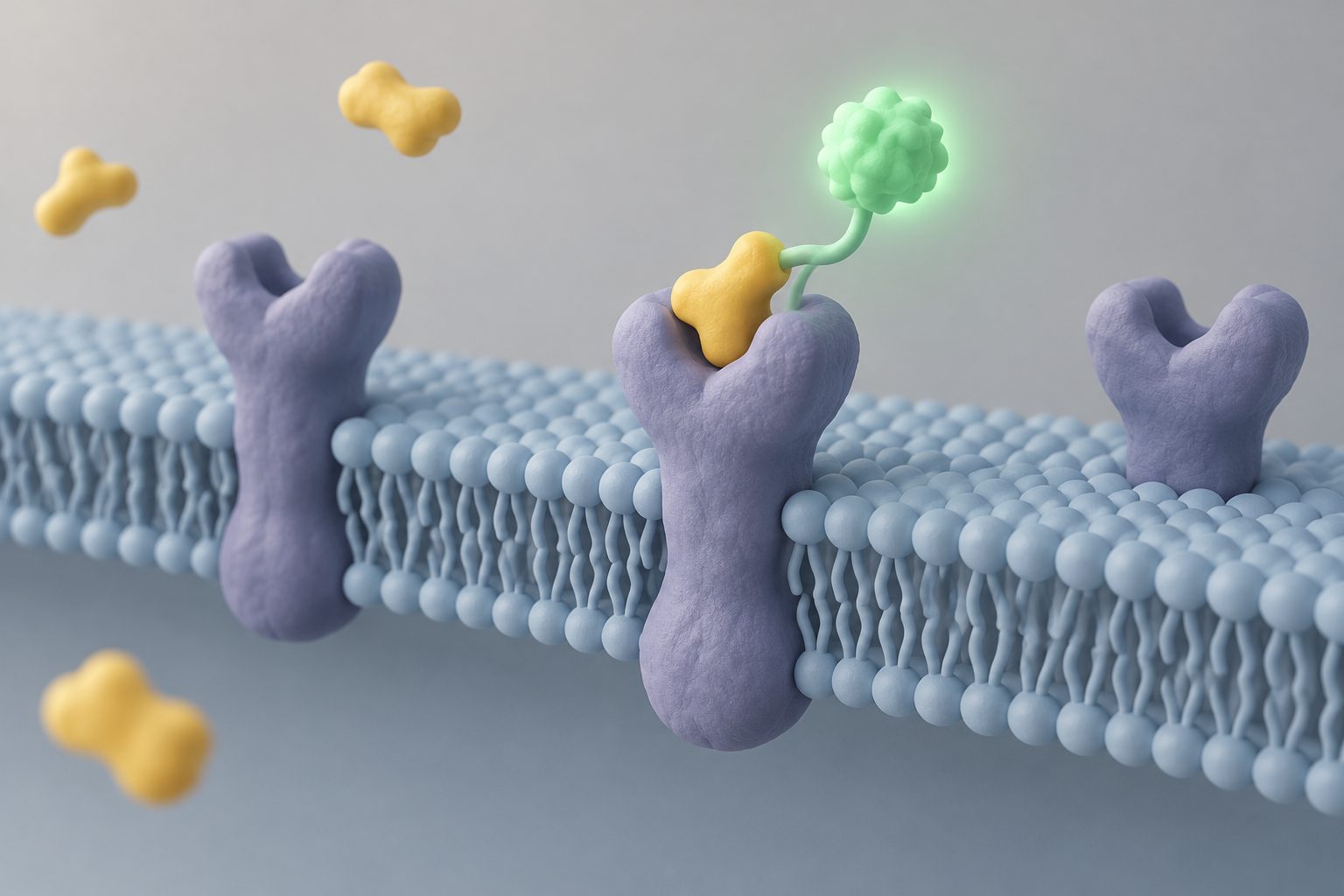
G-protein-coupled receptors, or GPCRs, are small cell-surface proteins whose shapes change when they bind different drugs. They have been difficult to study on their own with cryo-electron microscopy because they are too small and flexible, while commonly used markers can interfere with parts involved in signaling.
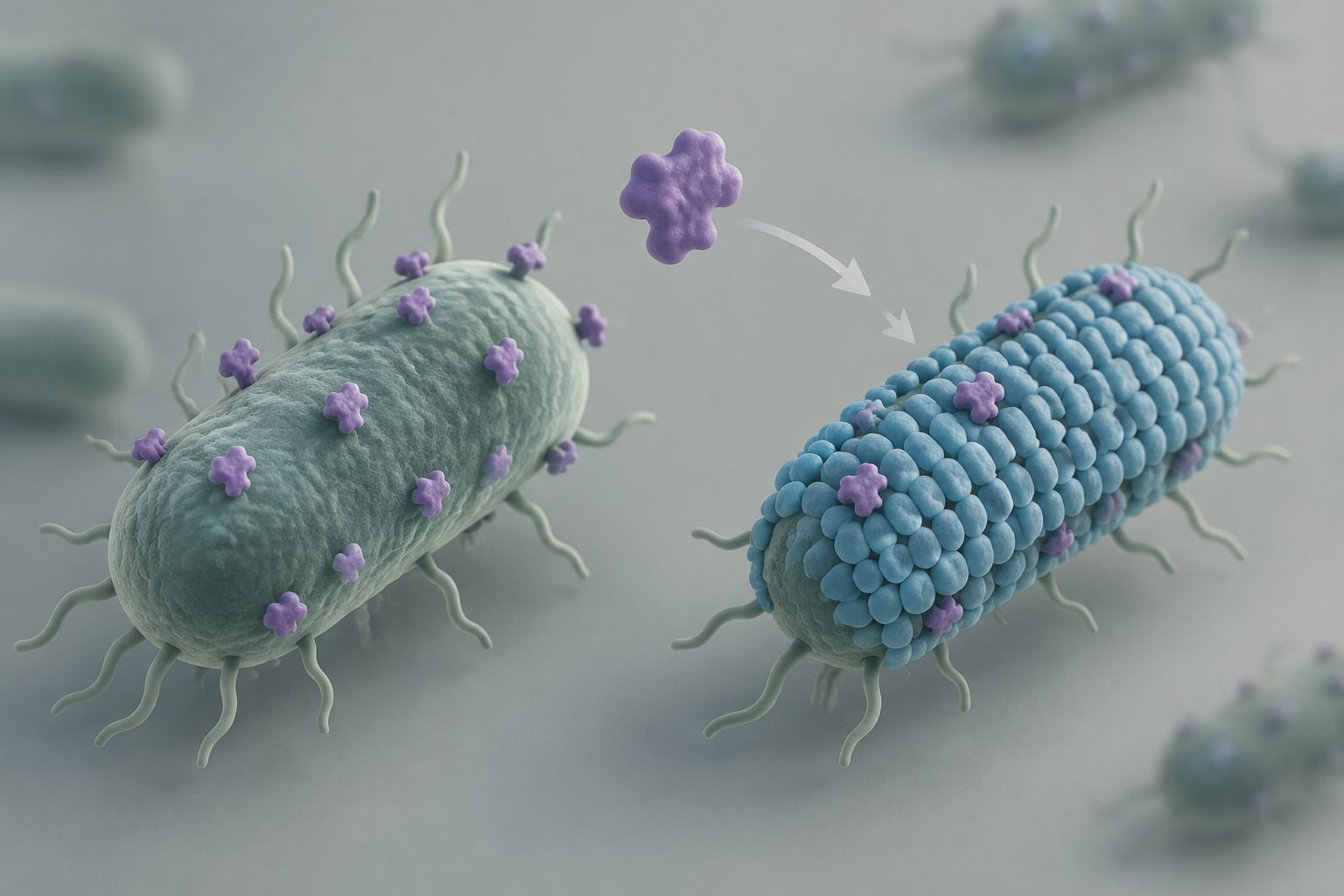
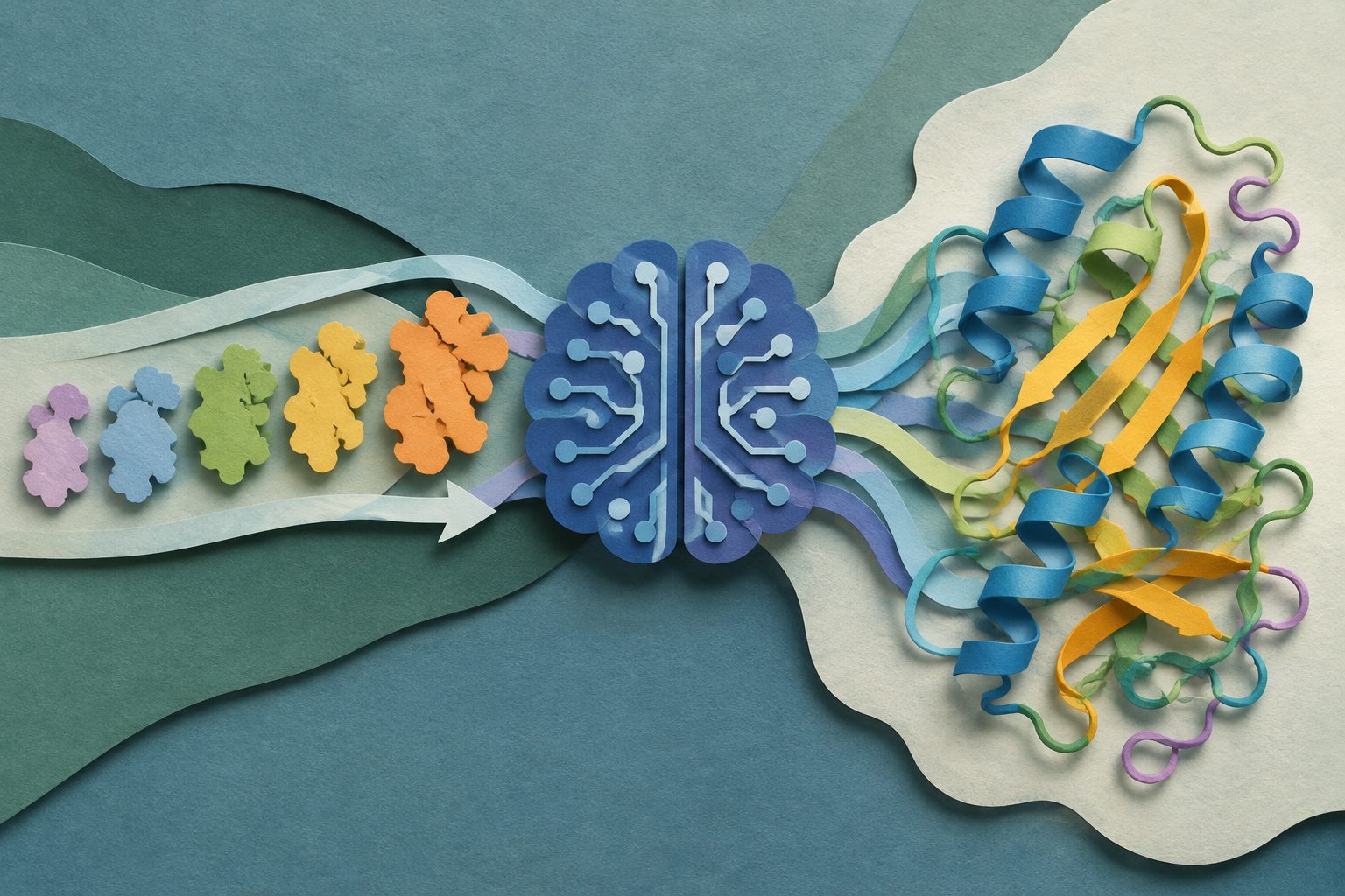
The researchers combined generative protein design with an artificial-intelligence method for predicting groups of possible protein shapes. They used the resulting markers to determine inactive-state structures for four drug-relevant GPCRs. They also attached a marker outside the beta-2 adrenergic receptor and observed changes in important intracellular parts of the receptor without a G protein present.
What the markers revealed
The researchers report that their design approach reliably produced rigid markers that could be attached at different sites on GPCRs, including difficult-to-use sites such as transmembrane helix 1. The markers supported high-throughput cryo-electron microscopy studies of inactive states in four pharmaceutically relevant GPCRs, allowing structural details related to each receptor’s pharmacology to be resolved.
For the beta-2 adrenergic receptor, an extracellular marker left the receptor’s intracellular vestibule with its native sequence and free of signaling partners. In the presence of a sufficiently potent agonist, the receptor showed an extensive rearrangement of ordered domains that resembled the form seen when it is bound to a G protein. The study also found that recent methods for predicting ligand-induced receptor shapes differed substantially from the observed structures.
Why the method matters
The markers provide a way to study GPCR shapes while leaving important intracellular signaling regions accessible and unoccupied. That could help researchers compare how full, partial and biased agonists alter receptor structure, rather than observing only receptors already locked into complexes with signaling partners or mimics.
The approach may make it easier to study other small, dynamic proteins and could support structural pharmacology and structure-based drug discovery. The researchers also suggest that receptor-alone structures could eventually help investigations of how GPCRs associate with signaling partners and larger signaling assemblies.
Evidence and caveats
The evidence comes from designed protein constructs and cryo-electron microscopy structures, including inactive-state structures for four GPCRs and ligand-bound structures of the beta-2 adrenergic receptor. The study directly characterized receptor rearrangements in the absence of a G protein, and compared the results with existing computational co-folding models.
The images may represent only some of the lower-energy states present for a given ligand. Higher-energy shapes may exist under conditions closer to those in living cells, and the freezing process is not instantaneous. The researchers also cannot rule out that the marker attached near transmembrane helix 1 altered the receptor’s range of shapes; they say this would need testing with other methods. Although the designs are expected to be transferable, extracellular markers may require further design for receptors with different extracellular pockets.