Brain differences in autism are highly individual, study finds
Across 1,344 brains, autistic people showed scattered cortical differences rather than one shared anatomical pattern.
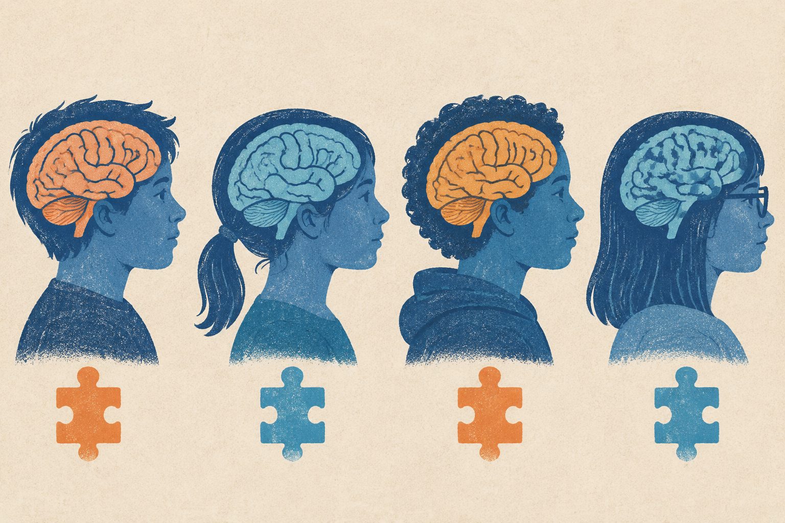
Editorial illustration — not from the study.
Researchers compared each person’s cortical organization with patterns expected from a large reference group. They found that autistic individuals had more extreme, spatially focused deviations than controls, but these differences were usually not concentrated in the same regions across people.
The changes tended to shift morphometric similarity—similarity based on several physical features of brain tissue—from association areas involved in higher-level processing toward sensory and visual areas. The researchers also identified a localized network of altered similarity, especially in occipital and temporal visual regions, and a continuous range of individual patterns linked to molecular maps and cognitive associations.
What the brain maps showed
Using data from 1,344 brains divided into 308 cortical regions, the researchers mapped how each autistic person’s brain differed from expected cortical organization. Autistic individuals had a higher prevalence of extreme, spatially focal deviations than controls, yet fewer than 12% of cases showed an extreme deviation in any single region.
The deviations followed broad features of the brain’s cortical hierarchy rather than appearing randomly. Morphometric similarity was preferentially redistributed from association and heteromodal cortex—areas involved in integrating information—toward sensory and visual territories. Network analysis highlighted a spatially localized subnetwork, dominated by occipital, temporal and visual regions.
Clustering of individual deviation maps revealed a reproducible, continuous axis with opposite overall patterns at its two ends. These patterns had different relationships with gene-expression and neurotransmitter-receptor maps, as well as different associations from cognitive meta-analyses.
Why varied patterns matter
The findings argue against treating autism as having one uniform cortical anatomy. Instead, they support a view in which autistic people can show different, individually focused departures from typical cortical organization, while those departures still follow broad cortical gradients and molecular patterns.
This subject-by-subject approach could help researchers study why autism has such varied biological and cognitive features. The authors describe it as a basis for mechanism-informed stratification, although the study does not establish that these brain patterns cause particular traits or symptoms.
Evidence and caveats
This is a multisite brain-imaging study using normative statistical models to compare each person’s cortical organization with expected patterns across a cohort of 1,344 brains. The analysis also compared the brain patterns with transcriptomic data, neurotransmitter-receptor density maps and cognitive meta-analytic associations.
The results show group differences and associations, not causes. The abstract does not provide details about the cohort’s demographic or clinical composition, and it does not establish how well the identified patterns predict an individual’s traits, diagnosis or future outcomes. Molecular and cognitive links based on mapped data also do not by themselves demonstrate a direct biological mechanism.
A study of illicit alcohol in two Ugandan cities found that alcohol strength was inconsistent and sometimes under-declared, reaching 44.2%. Some spirits contained methanol above safe limits, while opaque beers showed bacterial contamination and many production and retail sites had poor sanitation.
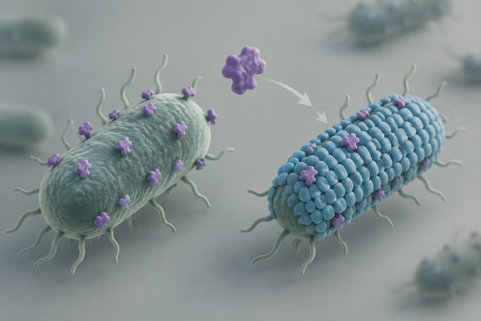
Researchers found that bacterial surface-layer proteins can evolve through modular changes and gene transfer, producing different outer structures. In closely related bacteria with the same type of surface layer, only pathogens showed resistance to the antimicrobial molecules LL-37 and lysozyme.
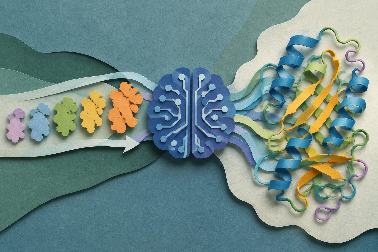
Researchers traced how AlphaFold 3 turns evolutionary differences among related protein sequences into three-dimensional structure predictions. They found that evolutionary diversity matters more than simply adding more sequences, helping explain both the system’s strengths and some of its failures.