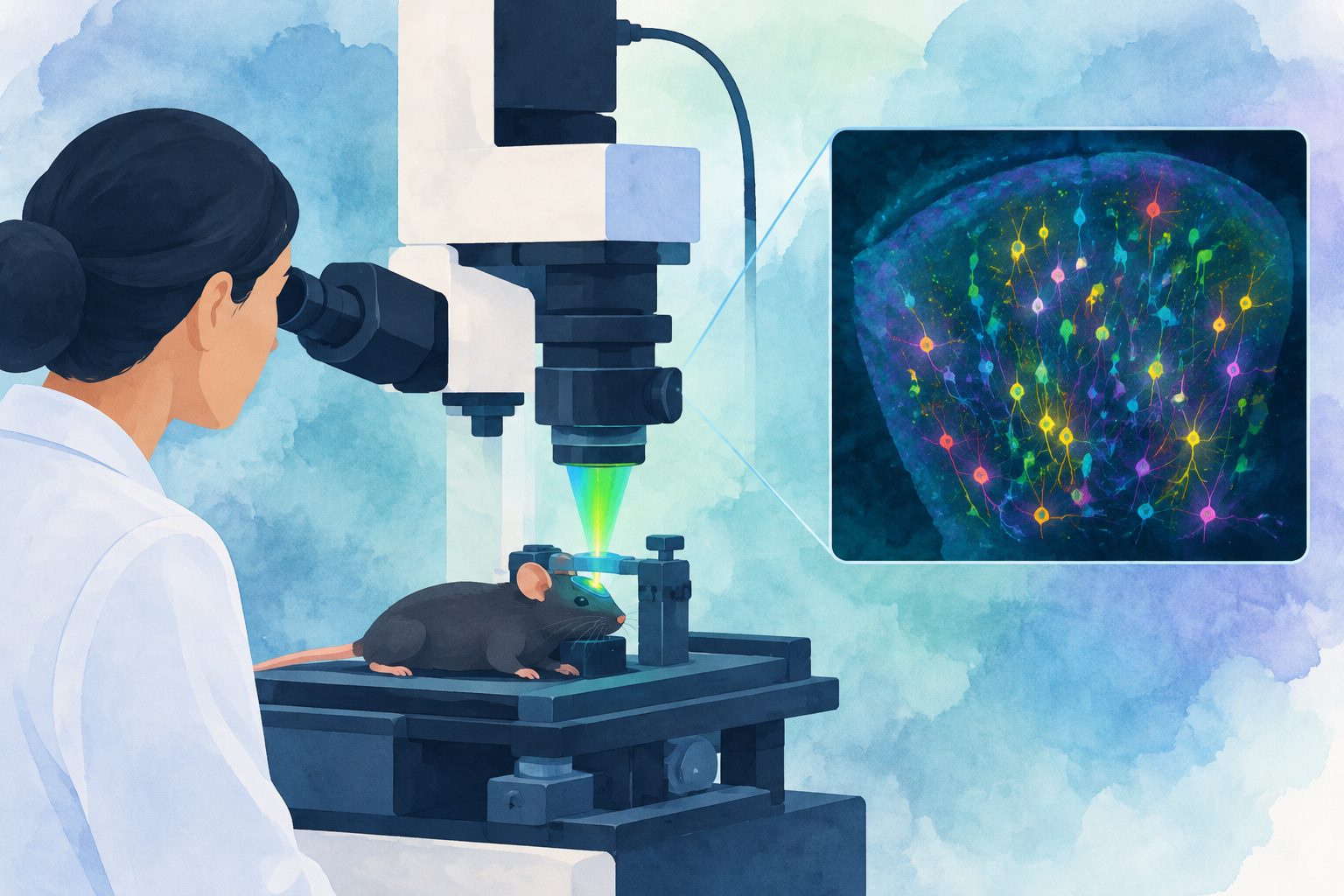
The system recorded activity across the hippocampus and nearby regions in behaving mice, and reached deep cortex in rats.
The microscope, called LIFT, combines a large imaging area with a custom scanning system and computer-based noise reduction. It recorded activity across the mouse hippocampus, surrounding white matter and nearby deep cortical layers, covering more than 1,500 cells at once in the intact brain.
In rats, the system produced population-level images with a strong signal 1.2 millimeters below the brain’s surface. The researchers say the microscope can be adjusted for different imaging areas and recording speeds, although those choices involve trade-offs.
Evidence and caveats
This was a technical demonstration using calcium imaging in two rodent species, including behaving mice. The microscope’s field of view, image detail and recording speed cannot all be maximized at once. In this study, available optical power limited imaging depth and signal quality, and the work focused on activity in neuronal cell bodies rather than finer cellular structures. The findings were demonstrated in mice and rats, not people.
// Source
Nature Communications · 2026 · DOI: 10.1038/s41467-026-78585-8
Authors: Hadas Frostig, Amy H. Monasterio, Hongjie Xia, Urvi Mishra, Siria Coello, Rebecca L. Suthard, Baldwin Britton, John T. Giblin, Steve Ramirez, Jerome C. Mertz, Benjamin B. Scott
Institutions: Boston University, Thorlabs (Germany), Thorlabs (United States)