X-ray maps how collagen fibres share loads inside spinal discs
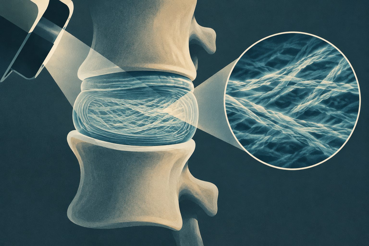
A new 3D imaging method links molecular changes in collagen fibrils to strains across intact mouse spinal discs.
Editorial illustration — not from the study.
The team combined 3D imaging with small-angle X-ray scattering to measure structural and mechanical properties from individual collagen fibres down to their nanoscale fibrils. The experiments were performed on intact mouse intervertebral discs before and after a 50-micrometre compressive displacement.
The measurements showed that the initial molecular stretch of fibrils varied in a layered pattern and was closely linked to strain in the larger fibres. Fibrils with greater pre-strain generally showed less strain under loading, a pattern consistent with load-sharing as their molecular structure unwinds.
How disc fibres share load
The imaging method, called TomoSAXS, produced three-dimensional maps connecting fibre-scale deformation with nanoscale changes in collagen fibrils. It reconstructed nanoscale measurements for more than 55% of the fibres in the samples.
A 2.2% reduction in disc height produced average compressive strains of 4.2 ± 3.3% in the outer disc, with local tissue strains above 15% near the vertebral endplates. At the fibre scale, highly curved fibres in the inner rear and rear-side regions of the outer disc reached strains of up to 20%.
The initial spacing between repeating features in collagen fibrils—a measure of molecular pre-strain—followed the disc’s layered structure and was strongly related to fibre strain. Greater initial pre-strain was associated with lower fibril strain and lower variation in fibril strain. Radial strain bridges and highly curved regions near the boundary between the disc’s outer ring and inner core also emerged as important sites for local load distribution.
Why the maps matter
The findings show how an intervertebral disc can coordinate mechanical behaviour across molecular, fibril, fibre and tissue scales while maintaining its shape and elasticity. Mapping these links in three dimensions could improve models of disc mechanics and provide a way to examine changes associated with ageing, disease or treatments.
The researchers also present TomoSAXS as a platform that could be applied to other biological and synthetic fibrous materials. The study demonstrates the imaging approach in intact mouse discs; its possible uses in therapies or material design were not tested here.
Evidence and caveats
This was an experimental imaging study of intact murine intervertebral discs, examined before and after a 50-micrometre compression at the Diamond Light Source synchrotron. The researchers combined three-dimensional phase-contrast imaging, displacement tracking and small-angle X-ray scattering, then used fibre orientations to separate signals from individual fibres.
The method recovered nanoscale properties for more than 55% of the fibres, rather than all fibres. The work also used a specific compression experiment in mouse tissue, so the results do not by themselves establish how human discs or other loading conditions behave. The proposed explanation involving molecular unwinding and load-sharing is consistent with the measured relationships, but the imaging study does not directly observe every molecular event.
Photographs taken over Braunschweig and Göttingen, Germany, during the 10–11 May 2024 magnetic superstorm showed red auroral rays extending into lower-altitude green rays. The researchers identify the diffuse green emission as a new type of feature, which they name Stable Auroral Green, or SAG, arcs.
A review in Physics Reports examines quantum bits built from semiconductor–superconductor devices. Their electrical tunability could support flexible quantum processors, but many of the proposed designs remain at the proof-of-concept stage.
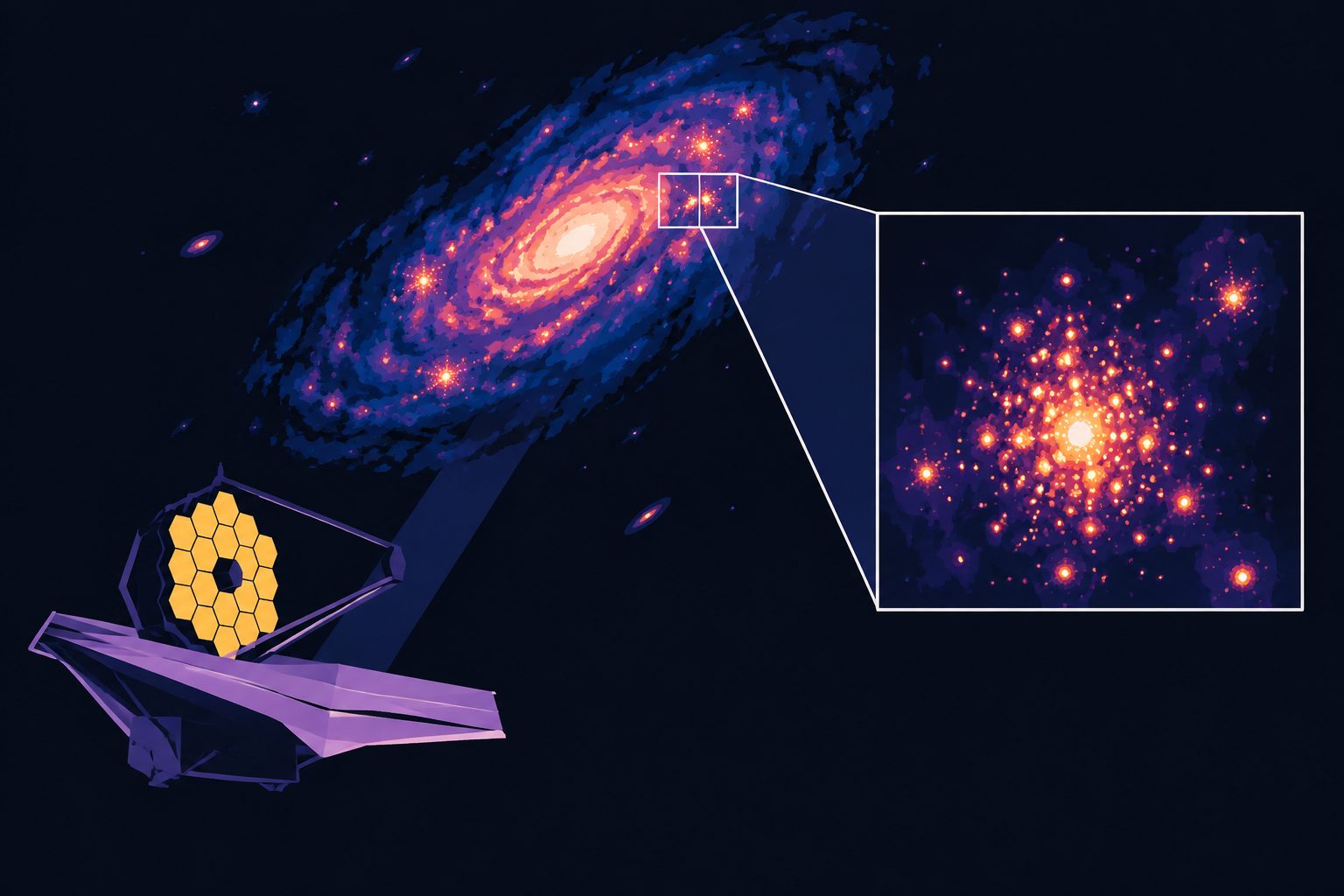
JWST observations have resolved three compact, young star clusters in the strongly lensed Cosmic Spear galaxy, seen as it was when the universe was young. The clusters are only a few million years old, yet appear massive, dense and gravitationally bound.