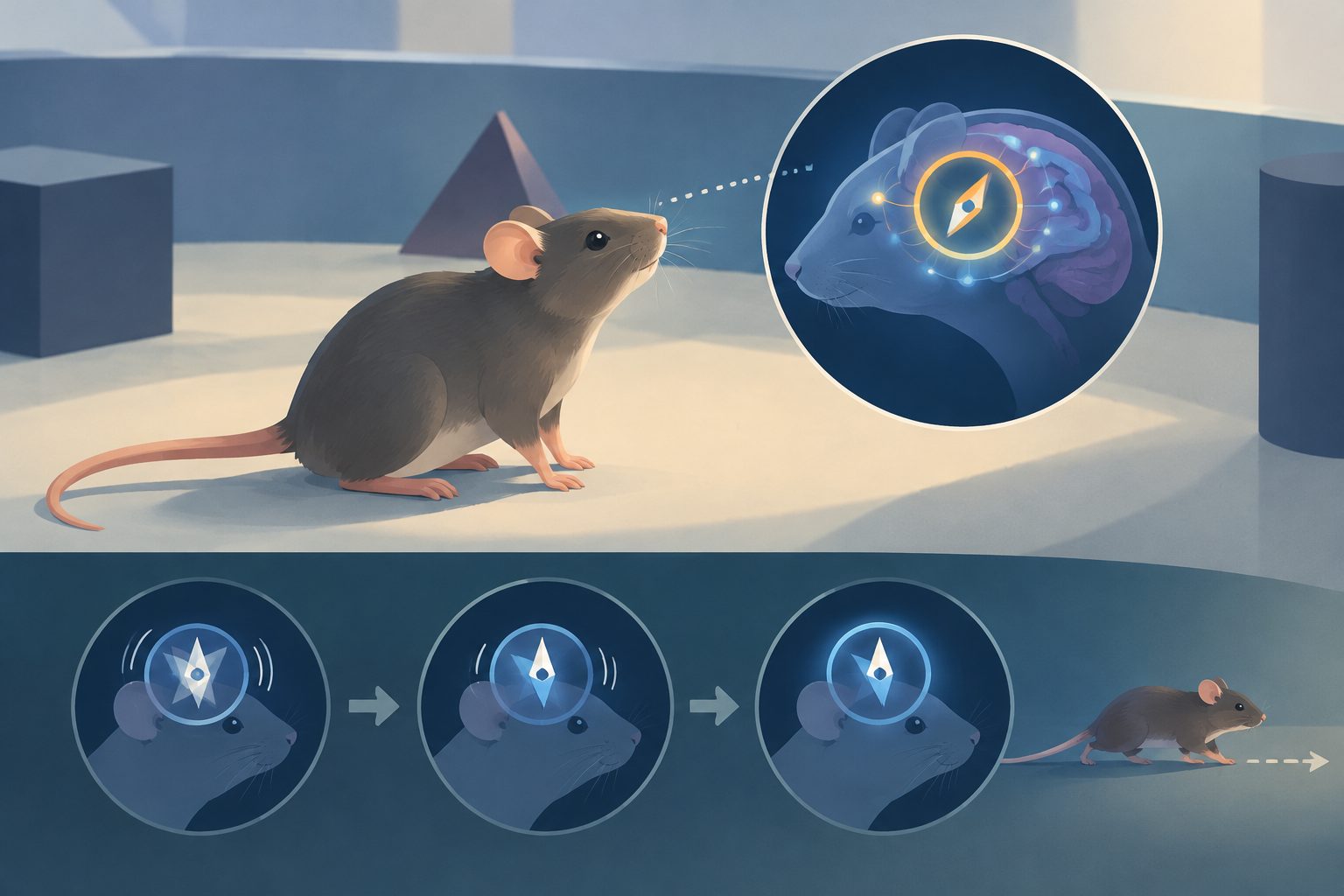
In head-fixed mice, brain signals for direction weakened during stillness but recovered hundreds of milliseconds before locomotion.
Researchers examined head-direction cells in the anterodorsal thalamus, a group of neurons involved in encoding which way an animal is facing. In head-fixed mice, restricting head movements disrupted the coordinated activity of these cells, even when individual cells could still respond to direction. The disruption was strongest during immobility and occurred in both real-world and virtual-reality settings.
When the mice began to run, the population activity returned toward its usual pattern. In one virtual-reality condition, this recovery began about 500 milliseconds before linear running started. A computational model reproduced similar changes by disrupting the connections between neighboring direction-sensitive neurons during stationary periods, supporting the idea that predicted movement helps maintain the directional signal.
How movement restored the compass
Complete head fixation, which creates a mismatch between expected and available vestibular signals, disrupted both individual-cell and population-level encoding of head direction. More selective restrictions on head-on-body movements impaired the coordinated activity of the head-direction population, while the effects were especially pronounced when the mice were immobile.
The researchers found that the network’s usual ring-shaped activity pattern was restored several hundred milliseconds before locomotion began. In virtual reality, the recovery preceded linear running by about 500 milliseconds, suggesting that an internal copy or prediction of an upcoming movement signal—not only feedback from movement already under way—helps restore the activity. When yaw-related vestibular input was removed entirely, this predictive stabilization was lost. A model in which connections among direction-sensitive neurons were shuffled during stationary periods reproduced key features of the experimental results.
Why the finding matters
The findings suggest that the brain’s internal compass is not simply a passive circuit that continuously integrates incoming sensory information. Its stability depends on the animal’s behavioral state and on signals related to forthcoming movement.
They also show that head fixation can change the neural code being studied, rather than merely holding the animal still. The results therefore point to a need for models of head-direction coding to include context, movement state and anticipatory motor signals when explaining how directional representations remain stable.
Evidence and caveats
The study combined recordings from head-direction neurons in head-restrained male mice during real-world and virtual-reality conditions with a computational model of a direction-coding network. The model reproduced changes seen in the experiments, but it does not by itself establish whether the stabilizing signal is a direct copy of a movement command or another internal prediction.
The results also depended on the sensory conditions: removing yaw input eliminated the predictive recovery, indicating that locomotion alone was not enough to maintain coherent activity in that setting. Head fixation additionally removed some natural head movements and, in virtual reality, pitch and roll signals, so the findings describe constrained experimental conditions rather than unrestricted navigation.