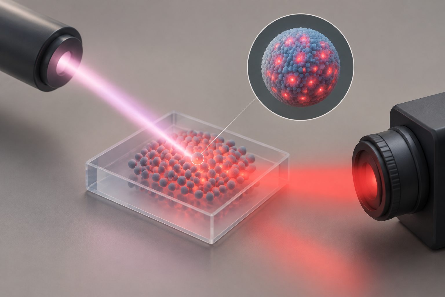
The team used holmium ions in core–shell nanoparticles to absorb 2-micrometre light and produce red light. Long-lived intermediate energy states in the holmium ions supported a four-photon upconversion process, with a relative upconversion-to-downconversion power conversion efficiency ratio of 13 ± 6%.
Materials & Energy1 day ago
Holmium nanoparticles turn 2-micrometre light into red light for imaging
The particles could let low-cost silicon detectors capture high-contrast images through highly scattering material.
How the particles convert light
The researchers report direct conversion of 2-micrometre shortwave infrared light to visible red light at 640 nanometres. The holmium ions acted as both the absorber and emitter, using their long-lived 5I7 and 5I6 intermediate states to support four-photon upconversion. Core–shell particles with well-passivated surfaces showed high-efficiency energy-transfer upconversion. Core-only particles instead displayed a surface-defect-mediated photon-looping and avalanche regime, which the researchers describe as previously unreported. The optimized core–shell particles were used for high-contrast, high-resolution shortwave infrared upconversion bioimaging through highly scattering media with low-cost silicon-based detection.
Why longer-wavelength light matters
Many conventional lanthanide-doped particles work with excitation wavelengths below 1700 nanometres. Extending upconversion to 2 micrometres could provide a way to use longer-wavelength infrared light in imaging while converting the signal to visible red light that silicon detectors can measure. The reported demonstration links this conversion to high-contrast bioimaging through scattering media and to relatively inexpensive detection hardware.
Evidence and open questions
This is a journal research article reporting measurements of holmium-doped nanoparticles and a bioimaging demonstration. The abstract gives a relative conversion ratio of 13 ± 6% and describes the imaging as high-contrast and high-resolution, but it does not provide absolute efficiency, imaging depth, biological subject details, or comparisons with existing imaging systems. It also does not establish how the particles perform in living organisms or in clinical use.
// Source
Journal of the American Chemical Society · 2026 · DOI: 10.1021/jacs.6c16249
Authors: Rakesh Arul, Zhao Jiang, Xinjuan Li, Fiona M. Bell, Alasdair Tew, Caterina Ducati, Akshay Rao, Zhongzheng Yu
Institutions: University of Cambridge