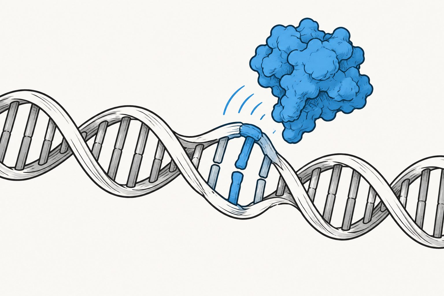
Tests of five DNA changes found structural and flexibility shifts extending beyond the damaged site.
Researchers examined DNA duplexes containing five modifications: a ribonucleotide, 8-oxoguanine, an abasic site, a nick and a one-nucleotide gap. Using single-molecule fluorescence measurements, they assessed changes in DNA bending, twisting and stretching at several positions around each modification.
All five modifications produced detectable differences from undamaged DNA, including the relatively small chemical difference between a ribonucleotide and a deoxyribonucleotide. Abasic sites, nicks and gaps caused the largest changes, and the effects extended to regions away from the damage itself.
How damaged DNA changed
Every tested modification changed the fluorescence signal compared with undamaged DNA. The measurements indicate that DNA damage alters the duplex’s structure and dynamics beyond the immediate lesion, including changes in bending, twisting, stretching and flexibility. Nicks and gaps produced the most pronounced shifts, while 8-oxoguanine, ribonucleotides and abasic sites caused subtler but still detectable changes.
The study used nine dye pairs for each DNA construct, with each measurement repeated three times. An automated analysis pipeline processed 162 measurements, allowing the researchers to compare the different damage types within the same experimental framework.
Why the shape matters
Repair proteins must find damaged sites among large stretches of undamaged DNA. The results support the idea that the DNA duplex itself may carry an early physical signal: damage could change the flexibility and overall shape of nearby DNA in ways that help guide repair-protein recruitment.
The findings also show that different lesions produce different structural effects. This may help explain why the nature and location of DNA damage can influence how it is recognized and repaired, although the study did not directly test protein recruitment.
Evidence and caveats
This was a laboratory study of specially prepared DNA duplexes, using confocal single-molecule fluorescence measurements and conformational modelling. The researchers compared five damage-related modifications with undamaged DNA and measured changes at multiple dye positions.
The fluorescence results show that distances and relative structural changes differ, but they do not provide a complete, absolute picture of DNA structure. The analysis commonly assumes that the fluorescent dyes can rotate freely enough for a standard orientation factor to apply; the researchers found some restriction of dye movement and could not rule out its contribution to uncertainty in absolute distance estimates. Direct time-resolved anisotropy measurements would be needed to assess that effect. The experiments also indicate a possible mechanism for repair-protein recruitment rather than demonstrating recruitment itself.
// Source
White Rose Research Online (University of Leeds, The University of Sheffield, University of York) · 2026
Open access versionPublished 2026-10-14 Authors: Sophie E. Fountain, Mahmoud A. S. Abdelhamid, D.M. Williams, Timothy D. Craggs
Institutions: University of Sheffield