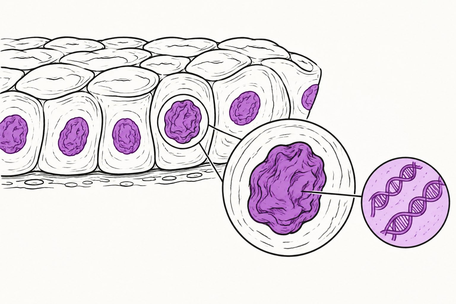
In cultured human cells, inward folds in the nuclear envelope helped accommodate changes in cell and nuclear shape.
Researchers used quantitative confocal imaging in 2.5D spheroids and controlled 2D cell layers to study nuclear wrinkles, which are inward folds of the nuclear envelope. Wrinkles were common in MCF10A cells but rare in hTERT-RPE1 cells, and their frequency increased as cultures became denser and nuclei became rounder.
Disrupting actin organization induced wrinkles, whereas disrupting microtubules did not. Live imaging showed that wrinkles could disappear and re-form at low cell density but became stable during prolonged confinement. The findings support a model in which excess nuclear surface folds as the nucleus changes shape, rather than wrinkles necessarily reflecting nuclear damage.
How nuclear wrinkles form
Nuclear wrinkles formed frequently in MCF10A epithelial cells but rarely in hTERT-RPE1 cells, showing strong cell-type differences. In MCF10A cultures, wrinkling increased with cell density and coincided with lower nuclear volume and cross-sectional area, along with a slight increase in nuclear height—changes consistent with nuclei becoming rounder.
Blocking actin filament organization robustly induced wrinkles in both cell types, while blocking microtubule organization did not. Acute cell rounding caused by trypsinization also produced widespread wrinkling across multiple cell types. Live-cell imaging showed that wrinkles were dynamic and reversible at low density but became stabilized under sustained confinement.
The researchers detected no nuclear-envelope rupture, DNA damage or stress-associated phosphorylation of histone H3 at serine 10 during wrinkle formation. Their quantitative analysis supports a passive geometric model: when nuclear shape changes, excess nuclear surface area can fold inward to accommodate that remodeling.
Evidence and caveats
The study combined quantitative confocal imaging of 2.5D spheroids and controlled 2D monolayers with live-cell imaging and short-term actin or microtubule disruption experiments. It examined several cultured human and other cell types, with especially detailed comparisons between MCF10A and hTERT-RPE1 cells.
The findings come from cell-culture models rather than intact tissues or organisms. The absence of envelope rupture, DNA damage and histone phosphorylation was based on the detection methods used, so it does not rule out every possible form of nuclear stress. Although the results support a passive geometric explanation, the experiments did not establish that cytoskeletal filaments never directly indent the nuclear envelope; the researchers note that some wrinkles overlapped with actin or microtubule signals, while others did not.