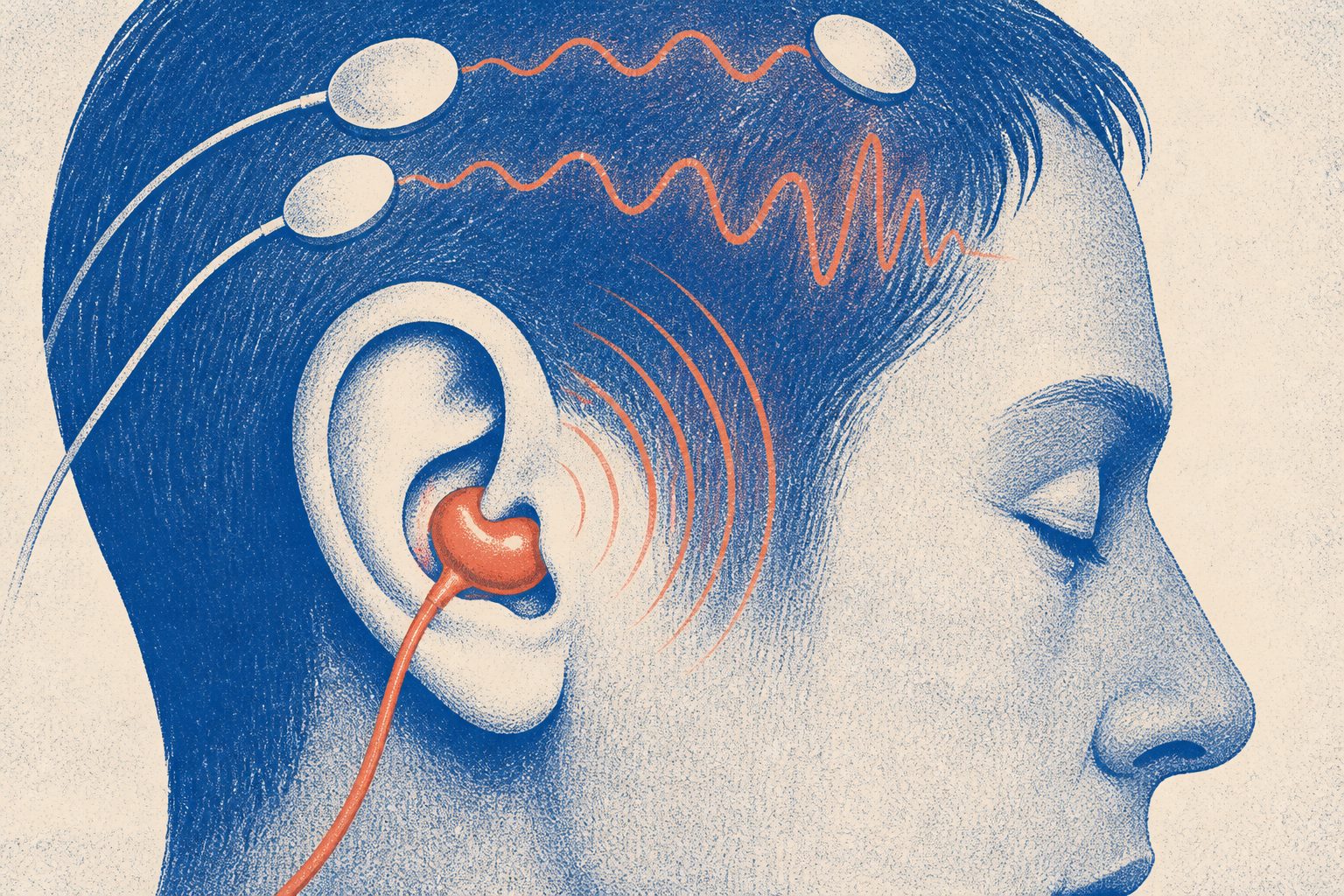
A dry earpiece made with silver conductive fabric produced signals that closely matched temporal scalp recordings, especially during sound-based tests.
The sensor was made from silver conductive fabric attached to an earpiece, with the aim of reducing the preparation and discomfort associated with conventional wet scalp electrodes. Participants completed eyes-open and eyes-closed recordings, a visual flicker task and auditory tasks while the in-ear and scalp electrodes recorded simultaneously.
The in-ear signals were weaker than recordings from some scalp locations, as expected for a sensor placed in the ear canal. However, the recordings were especially similar to temporal scalp signals during auditory steady-state response tests, suggesting that the sensor can capture activity associated with nearby temporal brain regions.
What the ear sensor recorded
In 15 healthy adults, the dry in-ear sensor recorded standard EEG responses during resting-state, visual and auditory tasks. Its alpha modulation ratio was lower than that measured with occipital scalp electrodes. Signals during the visual task were also attenuated compared with scalp recordings.
By contrast, the in-ear recordings were reported to be very similar to temporal scalp recordings, particularly during auditory steady-state response tests at 40, 55 and 70 Hz. The study reports a relationship between in-ear and temporal signals of r = 0.264, with p < 10⁻⁴. The researchers also report that the textile-based sensor required less preparation than conventional wet electrodes and was designed to be more flexible and comfortable for extended use.
Why an in-ear sensor matters
Scalp EEG can require skin preparation, conductive gel and lengthy setup, which can make prolonged monitoring uncomfortable or impractical. An in-ear sensor that records signals resembling those from a nearby temporal scalp site could offer a less intrusive way to collect EEG recordings.
The results support further development of wearable EEG devices, but they do not show that the sensor can replace standard scalp electrodes in clinical care or in every type of brain-monitoring application.
Evidence and caveats
This was a simultaneous recording study in 15 healthy adults, with the new sensor compared with wet electrodes at temporal and occipital scalp locations. The participants completed controlled resting, visual and auditory tasks, including auditory steady-state responses.
The study was small and did not test patients, long-term daily use or clinical monitoring. The in-ear signals were weaker for some measures, including the visual task, and the supplied materials do not report a direct, standardized measurement of comfort. The abstract describes the visual stimulation frequency as 12 Hz, while the methods excerpt describes a 15 Hz stimulus, so the reported protocol contains that inconsistency.