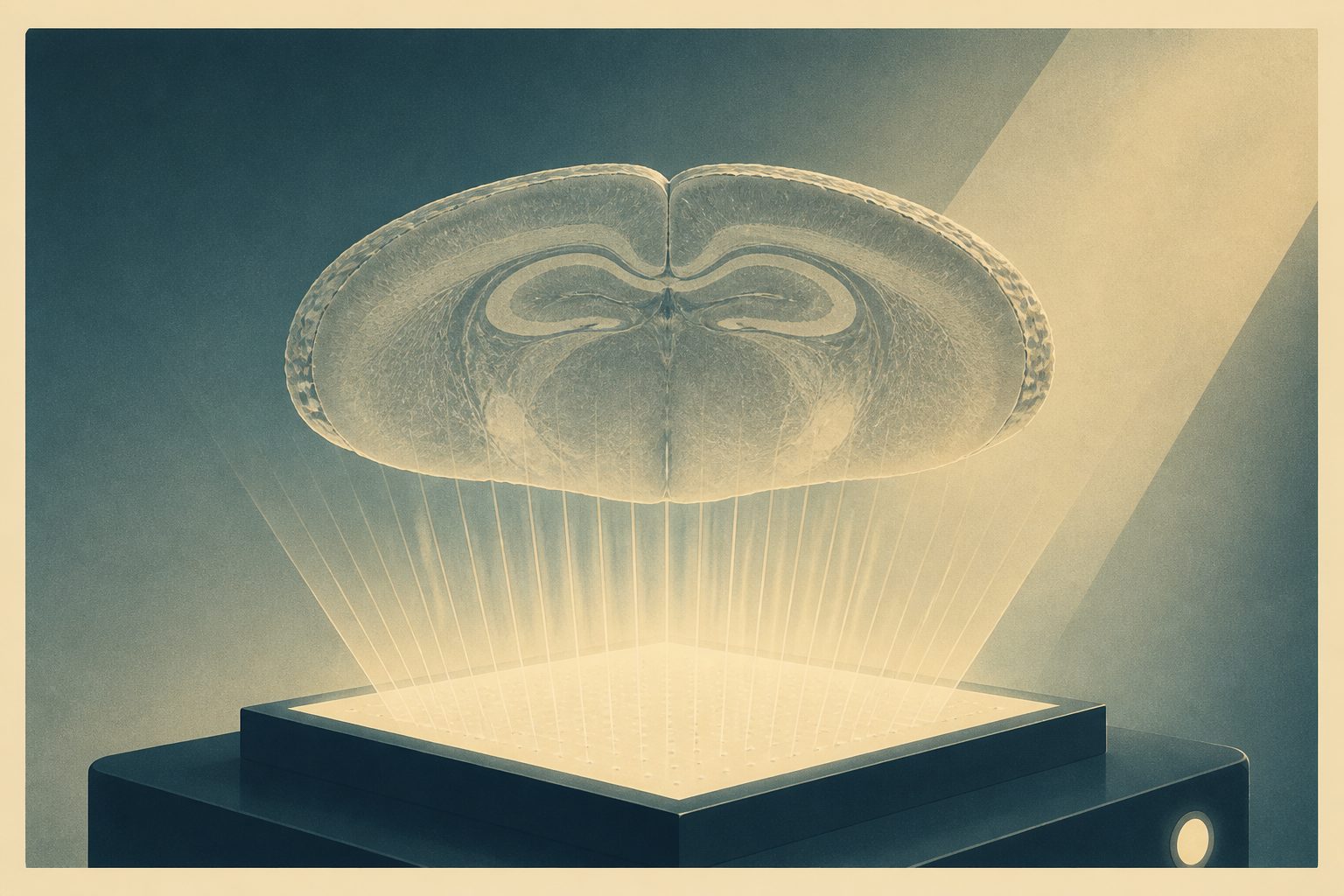
The system produced label-free three-dimensional images across a large area while accounting for light scattered through the tissue.
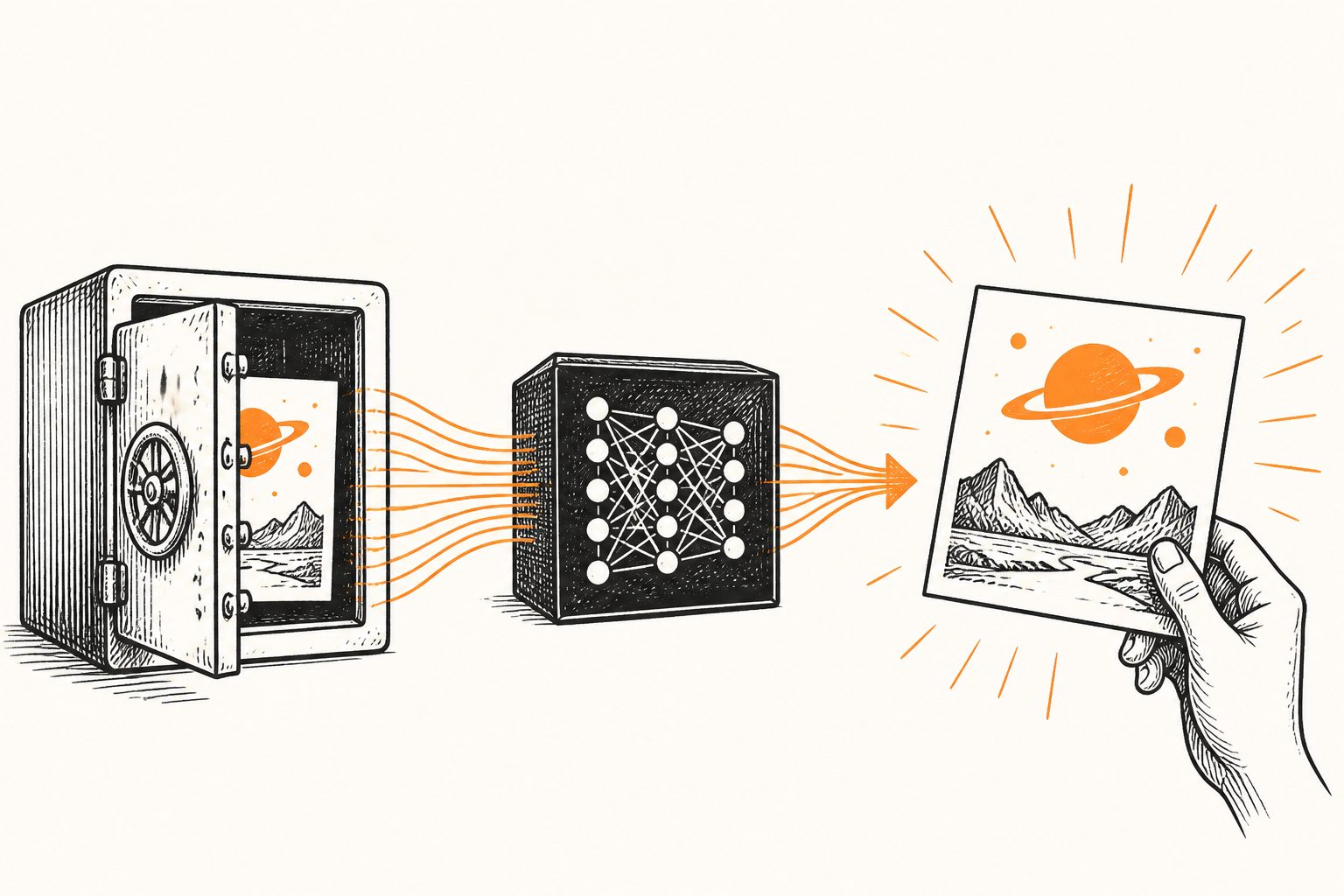
The method uses holograms recorded with several wavelengths and angled illumination, along with automatic calibration of the illumination angles. Its reconstruction process models multiple scattering—the redirection of light within a sample—rather than relying only on simpler first-order approximations.
In tests, the researchers reconstructed a custom multilayer polymer structure over 1.7 millimeters of depth and a 25-square-millimeter field of view. They also demonstrated on-chip, label-free imaging of an entire 500-micrometer-thick slice of optically cleared mouse brain.
Evidence and caveats
The evidence comes from an imaging-method study: a custom test structure was reconstructed under specified depth and field-of-view conditions, and the system was demonstrated on one type of biological sample described in the abstract. The abstract does not report the number of tissue slices, comparisons with a specific established microscope on biological samples, or quantitative accuracy measures for the mouse-brain images. The biological demonstration used an optically cleared tissue slice, so the results do not by themselves establish performance in intact, uncleared brain tissue or other samples.
// Source
Light Science & Applications · 2026 · DOI: 10.1038/s41377-026-02416-0
Authors: Mikołaj Rogalski, Julianna Winnik, Julia Dudek, Piotr Arcab, Emilia Wdowiak, Paweł Matryba, Marzena Stefaniuk, Piotr Zdańkowski, Maciej Trusiak
Institutions: Polish Academy of Sciences, Medical University of Warsaw, Warsaw University of Technology