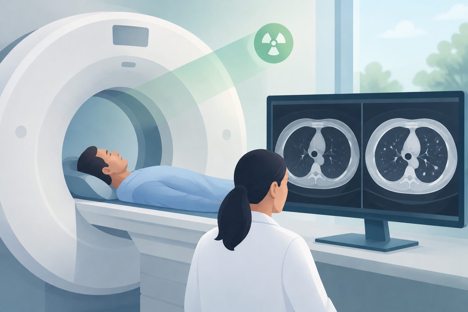
The woman arrived at an emergency department after severe overnight pain in the upper abdomen that had eased by the time she was assessed. Her examination and blood tests were not strongly specific for this condition. CT imaging identified an obstructed, enlarged cecum in the lesser sac, an abdominal space behind the stomach, with no reported signs of bowel-wall gas suggesting tissue injury.
Health & Medicine1 month ago
Researchers report a rare abdominal hernia in a patient with a brain fluid shunt
A 73-year-old woman was diagnosed by CT and underwent surgery after part of the bowel moved through an internal opening.
Low evidenceHuman studyInterpret with caution
Medical disclaimer: This article summarizes research findings and is for informational purposes only. It is not medical advice.
What was studied
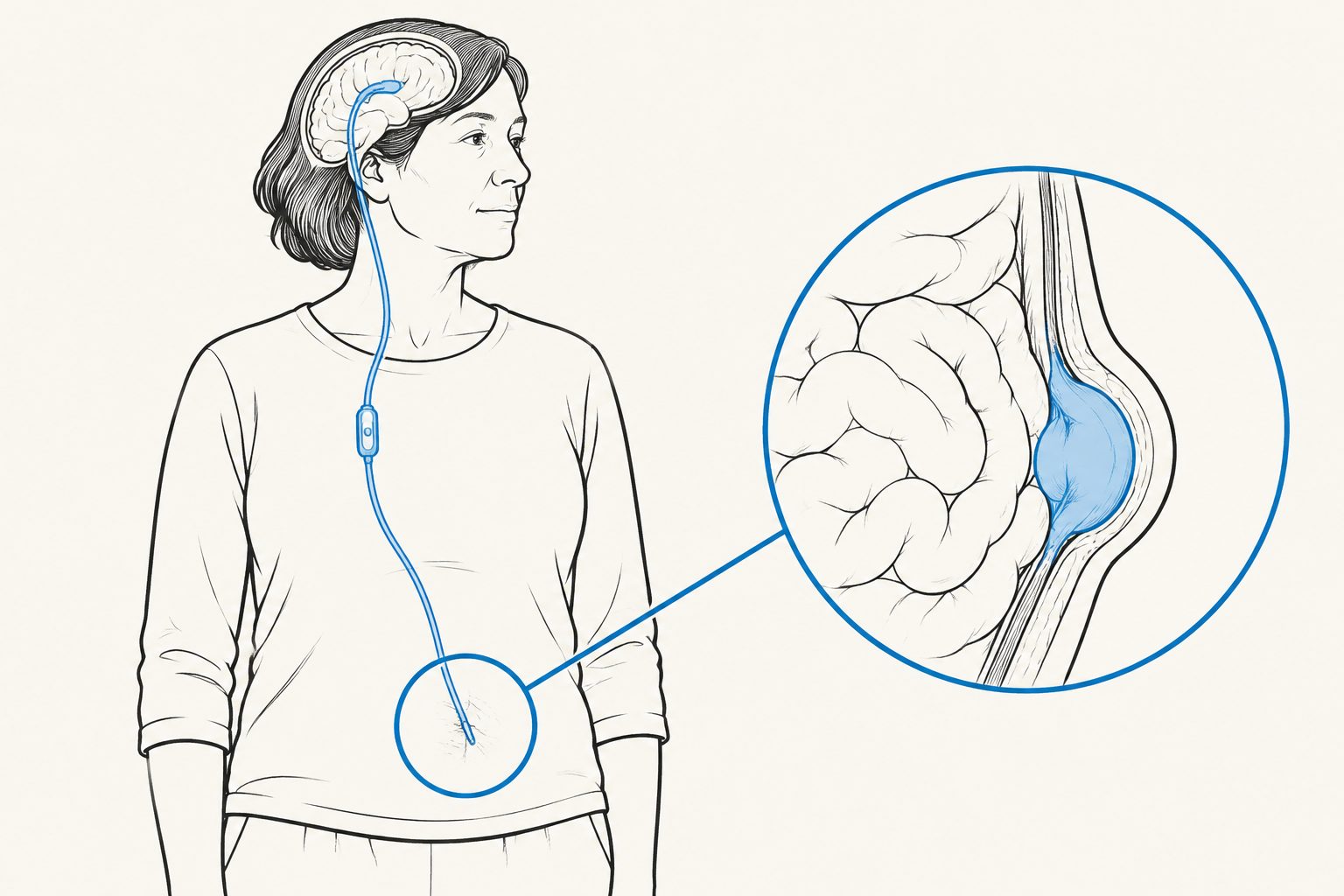
The article presents one human case and discusses previously published information about foramen of Winslow hernias. The patient was a 73-year-old woman who had received a ventriculoperitoneal shunt about four years earlier for hydrocephalus. The report describes her symptoms, examination, CT findings, attempted laparoscopic surgery, conversion to an open operation, and follow-up three weeks later. The excerpts do not provide a systematic-review method or a defined group of comparison patients.
Key observations
CT showed the cecum passing through the foramen of Winslow into the lesser sac and becoming obstructed and dilated. During surgery, the cecum and part of the small intestine were reduced; the cecum and nearby colon were swollen and redundant but still viable. The shunt catheter was seen in the abdominal cavity and was handled carefully during the operation. The patient was discharged on the fifth postoperative day according to the abstract, and the report describes an uncomplicated recovery at three-week follow-up. The article also states that there is no clear agreement on surgical management of these rare hernias or on abdominal surgery when a ventriculoperitoneal shunt is present.
Who this may apply to
The findings are most directly relevant as a description of one older adult with a foramen of Winslow hernia and a ventriculoperitoneal shunt. They should not be assumed to represent all people with abdominal pain, internal hernias, or shunts. The report does not determine whether the same symptoms, imaging findings, surgical difficulties, or recovery would occur in other patients.
Why this matters
The case illustrates that this rare internal hernia may have subtle or nonspecific symptoms and that imaging identified the problem in this patient. It also highlights an area where the authors report limited guidance: surgical management of the hernia and handling of a ventriculoperitoneal shunt during abdominal surgery. Because this is a single case, it does not establish how frequently this presentation occurs, whether the shunt contributed to the hernia, or which surgical approach is generally preferable.
Limitations & evidence assessment
The main limitation is that this is a single case report with no control group or comparison treatment. The follow-up reported is short, extending to three weeks after surgery, so longer-term outcomes are unknown. The condition is rare, and the authors state that its predisposing factors and several aspects of surgical management remain uncertain. Although the article includes a review of literature, the available excerpts do not describe a systematic search or formal evidence assessment, so the completeness and strength of that review are unknown.
Why this evidence level: This is a report of one human patient, not a controlled study. It describes a rare medical event and its management, so it can provide clinical context but cannot show how often outcomes occur or establish which approach is preferable.
Evidence levels are editorial estimates derived from study metadata — they are not clinical appraisals.
// Source
Journal of Case Reports and Images in Surgery · 2026 · DOI: 10.5348/100173z12pr2026cr
Authors: Prasenjit Raychaudhuri, Venkata V Raja Seelamanthula, Mohammed J Motamedi, Ammar Fadel, Allison A Elege, Rasika Hendahewa