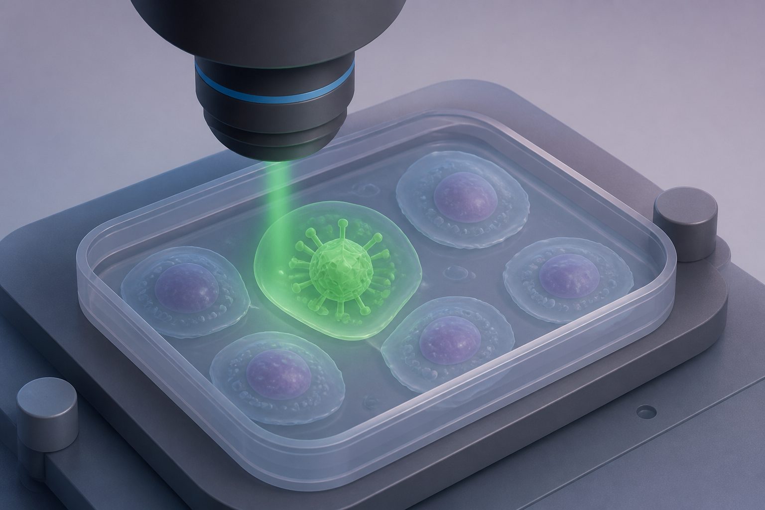
The method used cells’ own chemical signals to distinguish infected from uninfected cells without fluorescent tags.
The study applied optical photothermal infrared imaging and spectroscopy to living Spodoptera frugiperda Sf9 cells carrying no fluorescent reporter. Researchers examined cells 3, 6, 12 and 24 hours after baculovirus infection in a calcium fluoride sandwich setup.
Signals linked to proteins and nucleic acids provided spatial information that separated infected and uninfected cells. The measurements also captured differences between cells and showed how infection-related signals changed across the time points.
Chemical signals tracked infection
Imaging at a protein-associated band and a nucleic-acid-associated band distinguished infected from uninfected cells at each measured time point, reaching a balanced accuracy of 0.90. Point spectroscopy, which measures the chemical signal at specific locations, increased pairwise balanced accuracy to 0.94 and supported the biochemical basis of the image contrast. When the method classified cells across all four time points, overall accuracy was 0.71. Researchers also quantified differences between individual cells using a data-visualization method called UMAP, and used results across separate experimental replicates as an internal check on reproducibility.