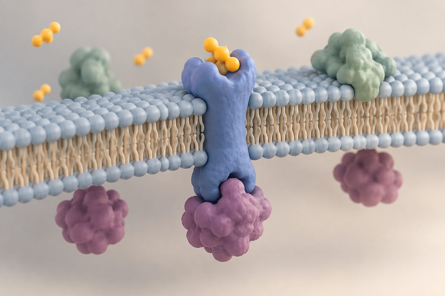
The method, called transmembrane PhoxID, uses a light-sensitive molecule attached to the outside of a transmembrane receptor. Light produces singlet oxygen, a reactive form of oxygen that can cross the cell membrane, allowing nearby proteins on both sides to be labelled for identification.
The researchers used the method on GRID2, GABAAR and GRM1 receptors in living mouse brains. The labelled proteins included components of transsynaptic nanocolumns at parallel fibre–Purkinje cell synapses, and the study identified a previously uncharacterized interaction between GABAAR and CAMKV in mouse and human brains.